. 2011 Mar-Apr;18(2):94-107.
doi: 10.1111/j.1399-3089.2011.00633.x.

Absence of Gal epitope prolongs survival of swine lungs in an ex vivo model of hyperacute rejection

Affiliations

Absence of Gal epitope prolongs survival of swine lungs in an ex vivo model of hyperacute rejection

Bao-Ngoc H Nguyen et al. Xenotransplantation. 2011 Mar-Apr.

Erratum in

  • Xenotransplantation. 2011 Jul-Aug;18(4):267

Abstract

Background: Galactosyl transferase gene knock-out (GalTKO) swine offer a unique tool to evaluate the role of the Gal antigen in xenogenic lung hyperacute rejection.

Methods: We perfused GalTKO miniature swine lungs with human blood. Results were compared with those from previous studies using wild-type and human decay-accelerating factor-transgenic (hDAF(+/+) ) pig lungs.

Results: GalTKO lungs survived 132 ± 52 min compared to 10 ± 9 min for wild-type lungs (P = 0.001) and 45 ± 60 min for hDAF(+/+) lungs (P = 0.18). GalTKO lungs displayed stable physiologic flow and pulmonary vascular resistance (PVR) until shortly before graft demise, similar to autologous perfusion, and unlike wild-type or hDAF(+/+) lungs. Early (15 and 60 min) complement (C3a) and platelet activation and intrapulmonary platelet deposition were significantly diminished in GalTKO lungs relative to wild-type or hDAF(+/+) lungs. However, GalTKO lungs adsorbed cytotoxic anti-non-Gal antibody and elaborated high levels of thrombin; their demise was associated with increased PVR, capillary congestion, intravascular thrombi and strong CD41 deposition not seen at earlier time points.

Conclusions: In summary, GalTKO lungs are substantially protected from injury but, in addition to anti-non-Gal antibody and complement, platelet adhesion and non-physiologic intravascular coagulation contribute to Gal-independent lung injury mechanisms.


Figures

Fig. 1
Fig. 1
Cumulative survival time of ex vivo perfused lungs. GalTKO, α galactosyl transferase knock-out pig lungs; WT, wild-type pig lungs; or hDAF+/+, hDAF transgenic pig lungs, were perfused with human blood; autologous, pig lungs from various sources perfused with autologous pig blood. Lung “survival time” represents the time of perfusion until which physiologic lung failure was determined, based on pre-defined criteria. #P = 0.0002 vs. WT; P = 0.178 vs. hDAF+/+; P = 0.0003 vs. autologous.
Fig. 2
Fig. 2
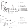
Pulmonary vascular resistance (PVR) and plasma levels of complement activation byproduct (C3a). (A) PVR expressed as a function of time for control and study groups. Time 0 represents measurements obtained during the first minute of lung perfusion, after circulation of the blood through the circuit for 10 min. PVR of GalTKO lungs was significantly lower than WT lungs and very similar to autologous lungs for the first 30 min. After 1 h, PVR of GalTKO lungs remained stable but higher than autologous lungs and then individual experiment levels usually rose sharply before failure. (B) Plasma C3a levels expressed as the amount of complement fragments produced above the pre-perfusion baseline (Δ). Complement activation was significantly curtailed in pig lungs lacking Gal. All data are shown as the mean±SEM of all surviving experiments in one group at each time.
Fig. 3
Fig. 3
Neutrophils (A) and monocytes (B) sequestration from circulation. Blood cell numbers measured serially by automated counting are expressed as percent remaining in circulation. Data expressed as change from the baseline and shown as the mean ± SEM of surviving experiments. *P < 0.05 vs. WT; #P < 0.05 vs. hDAF+/+.
Fig. 4
Fig. 4
Platelet and coagulation cascade activation profile. (A) Blood platelet counts measured serially by automated counting. Platelet sequestration was expressed as the percentage of platelets remaining in the perfusate at each time. (B) Platelet activation assessed by measuring the proportion of CD41+ platelets expressing CD62P by flow cytometry. (C) Platelet activation assessed by plasma levels of βTG released from platelet α granules. (D) Activation of the coagulation cascade was detected by the formation of thrombin measured by plasma levels of fragments F1 + 2. All data are expressed as change from the baseline (Δ) and shown as the mean ± SEM of surviving experiments. *P < 0.05 vs. WT; #P < 0.05 vs. hDAF+/+.
Fig. 5
Fig. 5
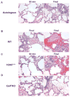
Histologic analysis of tissues from GalTKO and control perfused pig lungs. (A) An autologous control lung showed normal lung histology with thin alveolar septa and no pulmonary edema or intravascular thrombosis at 60 min. Mild cellular infiltration and endothelial cell activation is detected at elective termination after 300 min of perfusion. (B) WT lungs perfused with human blood showed severe interstitial/alveolar haemorrhage extended to the airway, cellular infiltration by polymorphonuclear granulocytes as well as severe intravascular thrombosis, in the cases illustrated at 13 and 21 min after reperfusion. (C) hDAF+/+ lungs showed largely preserved lung histology with thin alveolar septa, and limited congestion of capillaries and fibrin formation (not shown) at 60 min in a long survivor. In contrast, intravascular thrombosis and hemorrhage are prominent at lung failure (final, 165 min after reperfusion). (D) GalTKO lungs showed mild intravascular thrombosis, septal vascular congestion and edema at 60 min which were accentuated at lung failure (132 min after reperfusion).
Fig. 6
Fig. 6
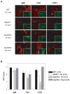
Two-color immunochemistry analysis of tissues from GalTKO and control perfused pig lungs. (A) Pig lung tissues collected at the indicated times were stained for IgM antibody deposition, classical complement activation fragment (C4d), or platelets (CD41) (red); co-staining for von Willebrand factor (VWF) identifies the endothelium (green insert). Strong staining of IgM on the endothelium in WT and hDAF+/+ groups was less prominent in GalTKO lungs at 10 min. Complement activation fragment C4d was associated with IgM deposits in capillaries, but much less frequently in large vessels. Both WT and hDAF+/+ lungs showed intense platelet deposition at 10 min. In contrast, platelet deposition was rare in GalTKO lungs at that 10 min time-point. However, at lung failure 60 min after reperfusion, there was prominent platelet deposition in association with alveolar septal endothelium in the GalTKO lungs. (B). Immunochemistry findings scored as the extent of tissue deposition as indicated in Methods. *There is a strong statistical trend towards decreased platelet deposition at 10 min in GalTKO lungs (P = 0.057 vs. WT). Original magnification × 200.
Fig. 7
Fig. 7
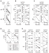
Anti-Gal and non-Gal antibody levels. (A–C) Antibody binding to Gal-sufficient (Gal+, A, B) or Gal-deficient (GalTKO, C) PAEC cells was measured by flow cytometry before and after perfusion of a Gal-sufficient (WT, A) or Gal-deficient (GalTKO, B,C) pig lung. Serum dilution was 1:10 for Gal+ cells and 1:4 for GalTKO cells. Results were expressed as MFI (median fluorescence intensity), or normalized MFI (MFI corrected for total immunoglobulins levels as indicated in Methods). (D) Anti-non Gal antibody levels were measured in a complement-dependent cytoxicity (CDC) assay using GalTKO peripheral blood mononuclear cells. Results were expressed as the percent lysis at 1:2 serum dilution. (E) Antibody levels to defined Gal and non-Gal carbohydrate antigens were measured by enzyme-linked immunosorbent assay and expressed as arbitrary units using a human serum pool as standard (1 A.U.). Arbitrary units were then normalized to total IgM or IgG levels as indicated in Methods. The percent change in antibody titer after lung perfusion for each condition is indicated as mean ± SD on each quadrant in panels A--D. The proportion of lungs showing decreased antibody levels is indicated in panel E. The legend indicates the individual GalTKO lungs subjected to ex vivo perfusion, ranked by increasing survival time (indicated in parenthesis), with assigned symbols and line characteristics.
Fig. 8
Fig. 8
Release of cytokines and other inflammatory mediators. Fold change in pig (A) and human (B) cytokines and IL8 chemokine, human platelet and coagulation activation markers (C), and monocyte-associated mediators (sCD14) and human neutrophil-(MPO) (D) measured at the time of lung failure in the perfused plasma by chemiluminescent protein array. Results were expressed as fold increase relative to the concentration of target protein pre-perfusion after normalizing the final concentration to total protein levels as described in Methods. Each symbol represents one individual GalTKO lung subjected to ex vivo perfusion. IL, interleukin; TNF, tumor necrosis factor alpha; IFNG, interferon gamma; sCD40LG, soluble CD40 ligand; SELP, soluble P-selectin; VWF, von Willebrand factor; F3, tissue factor; MPO, myeloperoxidase; sCD14, soluble CD14. #Human assays from Panel C cross-react with pig targets. The legend indicates the individual GalTKO lung perfusion experiments, ranked by increasing survival time (indicated in parenthesis), with assigned symbols characteristics. The dashed line represents no change in protein levels.

References

    1. Cooper DKC. Xenografting: how great is the clinical need? Xeno. 1993;1:25–26.
    1. Pierson RN., III Current status of xenotransplantation. JAMA. 2009;301:967–969. - PMC - PubMed
    1. Cooper DKC, Ye Y, Rolf JRLL, Zuhdi YYEN. The pig as potential organ donor for man. In: Cooper DKC, Kemp E, Reemtsma K, White DJG, editors. Xeno-Transplantation. Berlin: Springer-Verlag; 1991. pp. 481–500.
    1. Pierson RN, III, Dorling A, Ayares D, et al. Current status of xenotransplantation and prospects for clinical application. Xenotransplantation. 2009;16:263–280. - PMC - PubMed
    1. Pierson RN., III Antibody-mediated xenograft injury: mechanisms and protective strategies. Transpl Immunol. 2009;21:65–69. - PMC - PubMed

Publication types

MeSH terms