

High-resolution fMRI detects neuromodulation of individual brainstem nuclei by electrical tongue stimulation in balance-impaired individuals
- PMID: 21496490
- PMCID: PMC3105209
- DOI: 10.1016/j.neuroimage.2011.03.074
High-resolution fMRI detects neuromodulation of individual brainstem nuclei by electrical tongue stimulation in balance-impaired individuals
Abstract
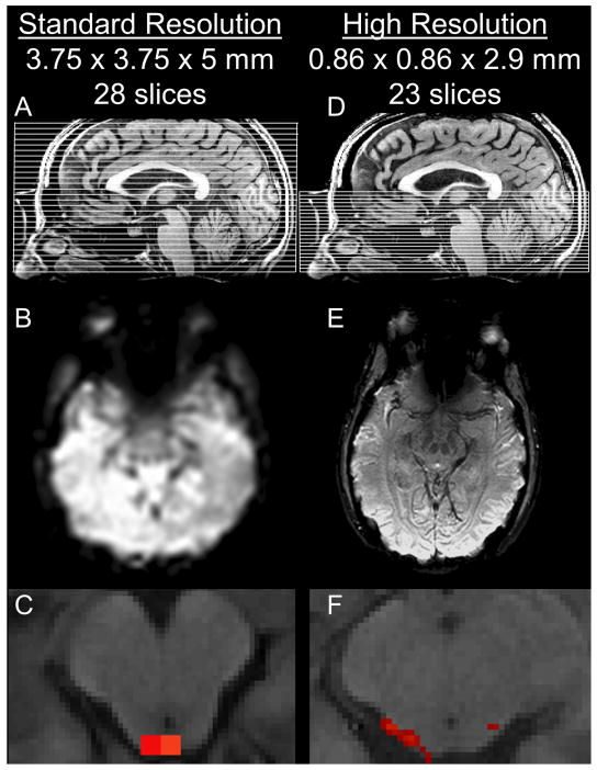
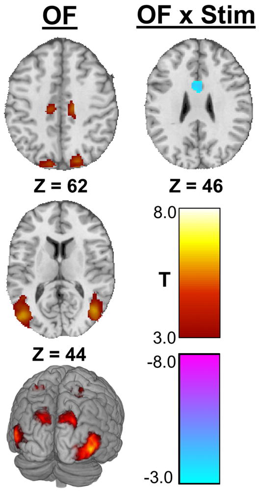
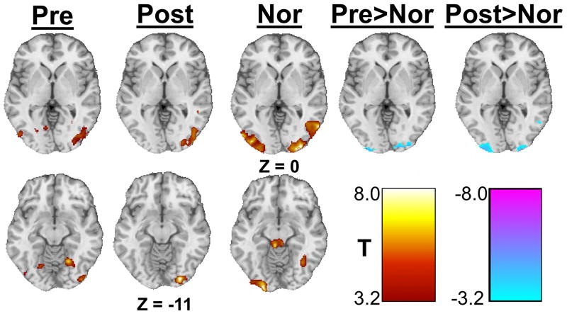
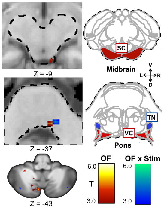
High-resolution functional magnetic resonance imaging (fMRI) can be used to precisely identify blood oxygen level dependent (BOLD) activation of small structures within the brainstem not accessible with standard fMRI. A previous study identified a region within the pons exhibiting sustained neuromodulation due to electrical tongue stimulation, but was unable to precisely identify the neuronal structure involved. For this study, high-resolution images of neural activity induced by optic flow were acquired in nine healthy controls and nine individuals with balance dysfunction before and after information-free tongue stimulation. Subjects viewed optic flow videos to activate the structures of interest. Sub-millimeter in-plane voxels of structures within the posterior fossa were acquired using a restricted field of view. Whole-brain functional imaging verified that global activation patterns due to optic flow were consistent with previous studies. Optic flow activated the visual association cortices, the vestibular nuclei, and the superior colliculus, as well as multiple regions within the cerebellum. The anterior cingulate cortex showed decreased activity after stimulation, while a region within the pons had increased post-stimulation activity. These observations suggest the pontine region is the trigeminal nucleus and that tongue stimulation interfaces with the balance-processing network within the pons. This high-resolution imaging allows detection of activity within individual brainstem nuclei not possible using standard resolution imaging.
Copyright © 2011 Elsevier Inc. All rights reserved.
Figures
Similar articles
-
Altered connectivity of the balance processing network after tongue stimulation in balance-impaired individuals.Brain Connect. 2013;3(1):87-97. doi: 10.1089/brain.2012.0123. Brain Connect. 2013. PMID: 23216162 Free PMC article.
-
Sustained cortical and subcortical neuromodulation induced by electrical tongue stimulation.Brain Imaging Behav. 2010 Dec;4(3-4):199-211. doi: 10.1007/s11682-010-9099-7. Brain Imaging Behav. 2010. PMID: 20614202 Free PMC article.
-
Electrical tongue stimulation normalizes activity within the motion-sensitive brain network in balance-impaired subjects as revealed by group independent component analysis.Brain Connect. 2011;1(3):255-65. doi: 10.1089/brain.2011.0029. Epub 2011 Sep 12. Brain Connect. 2011. PMID: 22433053 Free PMC article.
-
Neuroimaging and neuromodulation approaches to study eating behavior and prevent and treat eating disorders and obesity.Neuroimage Clin. 2015 Mar 24;8:1-31. doi: 10.1016/j.nicl.2015.03.016. eCollection 2015. Neuroimage Clin. 2015. PMID: 26110109 Free PMC article. Review.
-
The neuronal correlates of intranasal trigeminal function-an ALE meta-analysis of human functional brain imaging data.Brain Res Rev. 2010 Mar;62(2):183-96. doi: 10.1016/j.brainresrev.2009.11.001. Epub 2009 Nov 11. Brain Res Rev. 2010. PMID: 19913573 Free PMC article. Review.
Cited by
-
A feasibility study on the use of cranial nerve non-invasive neuromodulation to improve affected arm function in people in the chronic stage of a stroke.BMC Neurol. 2025 May 16;25(1):208. doi: 10.1186/s12883-025-04213-5. BMC Neurol. 2025. PMID: 40380081 Free PMC article. Clinical Trial.
-
Potential Mechanisms of Sensory Augmentation Systems on Human Balance Control.Front Neurol. 2018 Nov 12;9:944. doi: 10.3389/fneur.2018.00944. eCollection 2018. Front Neurol. 2018. PMID: 30483209 Free PMC article. Review.
-
Retention Effects of Long-Term Balance Training with Vibrotactile Sensory Augmentation in Healthy Older Adults.Sensors (Basel). 2022 Apr 14;22(8):3014. doi: 10.3390/s22083014. Sensors (Basel). 2022. PMID: 35459000 Free PMC article. Clinical Trial.
-
A Prospective, Multicenter Study to Assess the Safety and Efficacy of Translingual Neurostimulation Plus Physical Therapy for the Treatment of a Chronic Balance Deficit Due to Mild-to-Moderate Traumatic Brain Injury.Neuromodulation. 2021 Dec;24(8):1412-1421. doi: 10.1111/ner.13159. Epub 2020 Apr 29. Neuromodulation. 2021. PMID: 32347591 Free PMC article. Clinical Trial.
-
Using High Spatial Resolution to Improve BOLD fMRI Detection at 3T.PLoS One. 2015 Nov 9;10(11):e0141358. doi: 10.1371/journal.pone.0141358. eCollection 2015. PLoS One. 2015. PMID: 26550990 Free PMC article.
References
-
- Alvarez JC, Diaz C, Suarez C, Fernandez JA, Gonzalez del Rey C, Navarro A, Tolivia J. Aging and the human vestibular nuclei: Morphometric analysis. Mechanisms of Ageing and Development. 2000;114(3):149–172. - PubMed
-
- Anker AR, Ali A, Arendt HE, Cass SP, Cotter LA, Jian BJ, Tamrazi B, Yates BJ. Use of electrical vestibular stimulation to alter genioglossal muscle activity in awake cats. Journal of Vestibular Research. 2003;13(1):1–8. - PubMed
-
- Balaban CD, Thayer JF. Neurological bases for balance-anxiety links. Journal of Anxiety Disorders. 2001;15(1-2):53–79. - PubMed
-
- Barmack NH. Inferior olive and oculomotor system. Progress in Brain Research. 2006;151:269–291. - PubMed
MeSH terms
Grants and funding
LinkOut - more resources
Full Text Sources
Other Literature Sources
Medical
