. 2011 May;149(5):689-98.
doi: 10.1016/j.surg.2011.02.007.

Hands-free, wireless goggles for near-infrared fluorescence and real-time image-guided surgery

Affiliations

Hands-free, wireless goggles for near-infrared fluorescence and real-time image-guided surgery

Yang Liu et al. Surgery. 2011 May.

Abstract

Background: Current cancer management faces several challenges, including the occurrence of a residual tumor after resection, the use of radioactive materials or high concentrations of blue dyes for sentinel lymph node biopsy, and the use of bulky systems in surgical suites for image guidance. To overcome these limitations, we developed a real-time, intraoperative imaging device that, when combined with near infrared fluorescent molecular probes, can aid in the identification of tumor margins, guide surgical resections, map sentinel lymph nodes, and transfer acquired data wirelessly for remote analysis.

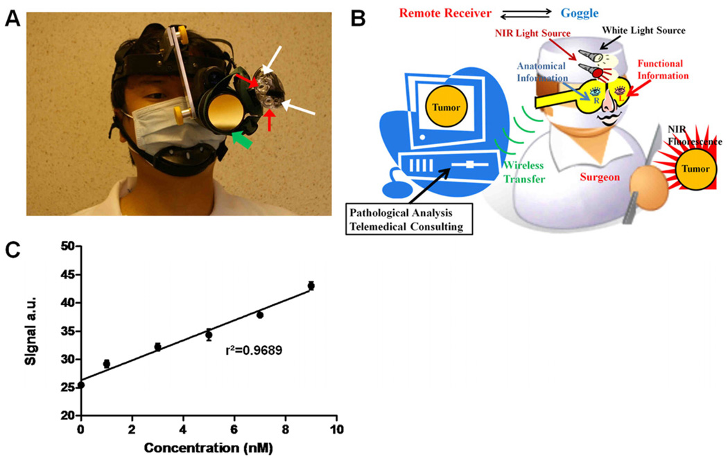
Methods: We developed a new compact, wireless, wearable, and battery-operated device that allows for hands-free operation by surgeons. A charge-coupled device-based, consumer-grade night vision viewer was used to develop the detector portion of the device, and the light source portion was developed from a compact headlamp. This piece was retrofitted to provide both near infrared excitation and white light illumination simultaneously. Wireless communication was enabled by integrating a battery-operated, miniature, radio-frequency video transmitter into the system. We applied the device in several types of oncologic surgical procedures in murine models, including sentinel lymph node mapping, fluorescence-guided tumor resection, and surgery under remote expert guidance.

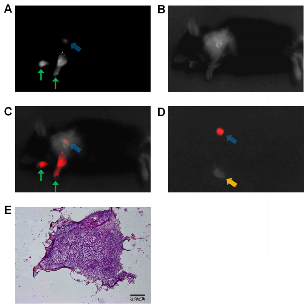
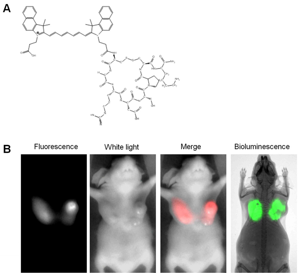
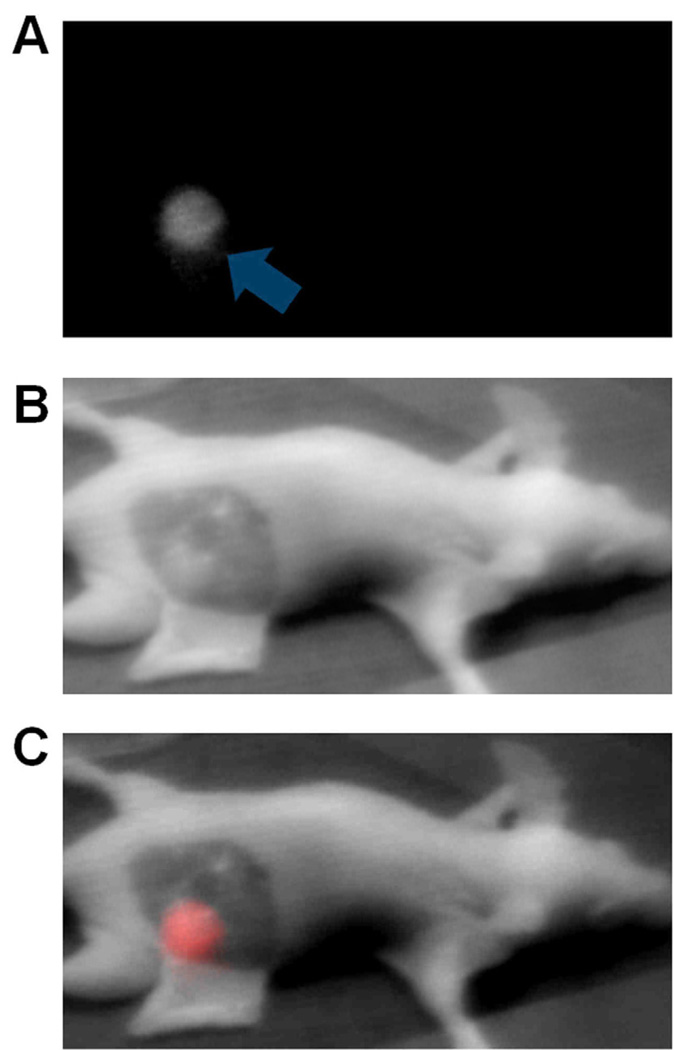
Results: Unlike conventional imaging instruments, the device displays fluorescence information directly on its eyepiece. When employed in sentinel lymph node mapping, the locations of sentinel lymph nodes were visualized clearly, even with tracer level dosing of a near infrared fluorescent dye (indocyanine green). When used in tumor resection, tumor margins and small nodules invisible to the naked eye were visualized readily. In a simulated, point-of-care setting, tumors were located successfully and removed under remote guidance using the wireless feature of the device. Importantly, the total cost of this prototype system ($1200) is substantially less than existing imaging instruments.

Conclusion: Our results demonstrate the feasibility of using the new device to aid surgical resection of tumors, map sentinel lymph nodes, and facilitate telemedicine.


Figures

Figure 1
Figure 1
Prototype intraoperative fluorescence imaging device. (A) Picture of the imaging device. Green arrows: detector; red arrows: NIR light sources; white arrows: white light sources. (B) Overview of the imaging system in a schematic diagram. Surgeon can capture functional information with one eye, while simultaneously obtaining anatomical information with the other eye. Real-time video can be transferred wirelessly to a remote site. (C) Sensitivity test of the device. NIR signal intensity versus indocyanine green concentration is plotted. Dots: mean values; error bars: standard deviation; r2: linear regression coefficient.
Figure 2
Figure 2
NIR sentinel lymph node mapping with the imaging system and indocyanine green. (A) NIR fluorescence image of a mouse at 5 minutes post-injection. Blue arrow indicates the putative sentinel lymph node. Green arrows indicate the injection points. (B) White light image of a mouse at 5 minutes post-injection, acquired under room light illumination. (C) NIR-white light merge image mice at 5 minutes post-injection. NIR fluorescence is pseudo colored in red and superimposed on white light image. The blue arrow indicates the putative sentinel lymph node at an axilllary position. Green arrows indicate the injection points (D) Ex vivo inspection of resected lymph nodes shown in NIR-light merge image. NIR fluorescence is psedocolored in red and superimposed on white light image. A blue arrow indicates the putative sentinel lymph node removed from an axillary position, while a yellow arrow indicates an ischiatic lymph node taken from the hindquarter as non-fluorescent control. (E) Histology of resected SLNs showing a representative image of a frozen section of SLNs with H&E staining.
Figure 3
Figure 3
NIR fluorescent probe LS 301 and tumor imaging (A) Structural illustration of LS 301. (B) Comparison of NIR fluorescence and bioluminescence images at 24 hours post-injection of LS 301. NIR fluorescence corresponds well with the bioluminscence in the tumor regions. NIR fluorescence and white-light images were acquired with the goggle device. Fluorescence was psedocolored in red in the fluorescence-white light merge image. Bioluminescence image and radiograph were captured with Carestream In-Vivo Multispectral FX PRO imaging system.
Figure 4
Figure 4
Intraoperative tumor imaging with goggle device and LS 301. (A) Images of a mouse at 24 hours after intravenous injection of LS 301. NIR fluorescence (Left), White light (middle), and NIR-white light merge (right) images are shown. Major nodules in both breasts as well as small nodules can be clearly identified. Green arrows: major tumor nodules; Purple arrow: small tumor nodule. (B) Images of a mouse during tumor resection. Tumor margins emit high florescent signal, as shown in the bright “rim” of left tumor. Several residual tumors and small nodules that are non-obvious to naked-eye are also identified. Blue arrows: tumor margins at cross-section of a major nodule; Purple arrow: small tumor nodule; Grey arrow: liver; Green dotted circle: residual tumor intentionally left from previous resection. (C) Ex vivo fluorescence, white light and merged images of tissues from dissected organs: 1.heart, 2.lung, 3.spleen, 4.kidney, 5.liver, 6.skin, 7. muscle, 8.brain, 9.tumor 10. blood. Resected tumor tissue shows high fluorescent signal (Green dotted circle). Liver and kidney also exhibit high fluorescence level as they are the major organs responsible for excretion and metabolism of imaging agents. (D) Fluorescence and color microscopy of resected tumor tissues. Merged autofluorescence (480ex/535em, green) and LS301 NIR fluorescence (775ex/810em, red) (left) along with color microscopy after H&E staining of the same section (right). High NIR florescence from LS 301 was found in the tumor, as well as on the margin, as shown in red color on the left image. Yellow arrows: tumor proper; Blue arrows: tumor margin.
Figure 5
Figure 5
Fluorescence-guided surgery images acquired via wireless transfer. Real-time video was transferred to remote computer using a RF transmitter/receiver set. (A) NIR fluorescence image, (B) white light, and (C) merged image of mouse 24 hours post-injection demonstrating the high signal from tumor tissue prior to resection.

References

    1. Collins L, et al. Outcome of Women with Ductal Carcinoma In Situ (DCIS) Treated with Breast-Conserving Surgery Alone: A Case-Control Study of 225 Patients from the Cancer Research Network. Modern Pathology. 2009;22:34A–35A.
    1. Vicini FA, et al. Impact of young age on outcome in patients with ductal carcinoma-in-situ treated with breast-conserving therapy. Journal of Clinical Oncology. 2000;18:296–306. - PubMed
    1. Albo D, et al. Anaphylactic reactions to isosulfan blue dye during sentinel lymph node biopsy for breast cancer. American Journal of Surgery. 2001;182:393–398. - PubMed
    1. Amr D, Broderick-Villa G, Haigh PI, Guenther JM, DiFronzo LA. Adverse drug reactions during lymphatic mapping and sentinel lymph node biopsy for solid neoplasms. American Surgeon. 2005;71:720–724. - PubMed
    1. Aydogan F, Celik V, Uras C, Salihoglu Z, Topuz U. A comparison of the adverse reactions associated with isosulfan blue versus methylene blue dye in sentinel lymph node biopsy for breast cancer. American Journal of Surgery. 2008;195:277–278. - PubMed

Publication types