Review
. 2011 Jun;21(3):296-305.
doi: 10.1016/j.sbi.2011.03.009. Epub 2011 Apr 14.

Topological constraints: using RNA secondary structure to model 3D conformation, folding pathways, and dynamic adaptation

Affiliations
Review

Topological constraints: using RNA secondary structure to model 3D conformation, folding pathways, and dynamic adaptation

Maximillian H Bailor et al. Curr Opin Struct Biol. 2011 Jun.

Abstract

Accompanying recent advances in determining RNA secondary structure is the growing appreciation for the importance of relatively simple topological constraints, encoded at the secondary structure level, in defining the overall architecture, folding pathways, and dynamic adaptability of RNA. A new view is emerging in which tertiary interactions do not define RNA 3D structure, but rather, help select specific conformers from an already narrow, topologically pre-defined conformational distribution. Studies are providing fundamental insights into the nature of these topological constraints, how they are encoded by the RNA secondary structure, and how they interplay with other interactions, breathing new meaning to RNA secondary structure. New approaches have been developed that take advantage of topological constraints in determining RNA backbone conformation based on secondary structure, and a limited set of other, easily accessible constraints. Topological constraints are also providing a much-needed framework for rationalizing and describing RNA dynamics and structural adaptation. Finally, studies suggest that topological constraints may play important roles in steering RNA folding pathways. Here, we review recent advances in our understanding of topological constraints encoded by the RNA secondary structure.


Figures

Figure 1
Figure 1
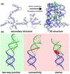
Topological constraints encoded by the RNA secondary structure. (a) Secondary and tertiary structure of group IIC intron from O. iheyensis (PDB 3IGI) [57]. (b) Connectivity and steric constraints in a two-way junction.
Figure 2
Figure 2
Topological constraints confine the allowed inter-helical orientations across bulges. (a) Three inter-helical Euler angles (αhβhγh) used to define the relative orientation of 5' and 3' helices across two-way junctions containing X and Y (X ≥ Y) single stranded (S) residues. (b) 3D inter-helical orientation maps showing individual 2D projections along each plane of a 3D cube with associated correlation coefficients (R) between the inter-helical twist angles for three-nucleotide bulged RNA TAR (S3S0, HIV-1 TAR). The ligand-bound, NMR-RDC, NMR-MD, PDB-derived, and topologically computed inter-helical distributions are shown in black, yellow, blue, red, and grey, respectively. (c) The PDB-derived inter-helical distributions for various families of Y-strand length junctions.
Figure 3
Figure 3
Determining backbone global conformation of (a) tRNAAsp and (b) Tetrahymena ribozyme P4–P6 domain based on secondary structure constraints and limited tertiary contacts. Predicted structures (in red) are superimposed onto the native X-ray structure (in blue). Adapted from [34•] and [29•] and (Flores, Sherman, Bruns, Eastman, and Altman, Transactions on Computational Biology and Bioinformatics, 2010, in press). Secondary structure motifs within green boxes correspond to motif environments that were subjected to constraints derived from comparison to independent crystal and NMR structures. Blue bars between bases correspond to imposed stacking interactions and red connectors denote imposed tertiary contact constraints.
Figure 4
Figure 4
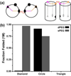
Folding simulations of model HJH motifs demonstrate the formation of tertiary contacts is selected based on contact location linker – single PEG linker (sPEG) and double PEG linkers (dPEG) – properties. (a) Schematic representation of the sPEG HJH motif in a side-by-side helical configuration. Tertiary contacts, highlighted by diamond, circle, and triangle, were placed in three separate locations and investigated computationally. (b) Comparison of the fraction folded sPEG (grey) and dPEG (black) in 1 M monovalent salt concentration for three tertiary contact. The two junction topologies have different conformational preferences (sPEG prefers the diamond position, dPEG prefers circle) and folding specificities (sPEG is more receptive to tertiary contact location) which results in differences of folded fractions. Adapted from [13••].
Figure 5
Figure 5
Formation of helical elements in the preQ1 riboswitch. All-atom Go model folding simulations of the preQ1 riboswitch showing formation of the P1 (red) and P2 (blue) helix contacts as a function of the total native contacts in the presence and absence of preQ1 ligand. Results are averaged over 78 kinetic folding simulations. Adapted from Feng et al [51].
Figure 6
Figure 6
Size-encoded adaptation of RNA by conformational selection using small molecules. (a) Inter-helical orientation maps of small-molecule bound RNA crystal structures for HIV DIS kissing dimers (2FCX, 2FCY, 2FCZ, 2FD0) according to solvent accessible surface area (SAS) of the ligand. Predicted junction-allowed topological distributions (in grey) for corresponding two-way junctions of type 2–1 in the “free” state are shown in the cube plot. Shown to the right are correlation plots between each small molecule’s SAS and the corresponding bound RNA’s inter-helical angles (αhβhγh) and Kd. (b) X-ray structures of two-way RNA junction from a are shown bound to their small molecules in order of increasing size. Aminoglycosides are shown in red.

Similar articles

Cited by

References

    1. Murthy VL, Srinivasan R, Draper DE, Rose GD. A complete conformational map for RNA. J Mol Biol. 1999;291:313–327. - PubMed
    1. Musselman C, Pitt SW, Gulati K, Foster LL, Andricioaei I, Al-Hashimi HM. Impact of static and dynamic A-form heterogeneity on the determination of RNA global structural dynamics using NMR residual dipolar couplings. J Biomol NMR. 2006;36:235–249. - PubMed
    1. Riordan FA, Bhattacharyya A, McAteer S, Lilley DM. Kinking of RNA helices by bulged bases, and the structure of the human immunodeficiency virus transactivator response element. J Mol Biol. 1992;226:305–310. - PubMed
    1. Lilley DM, Clegg RM, Diekmann S, Seeman NC, Von Kitzing E, Hagerman PJ. A nomenclature of junctions and branchpoints in nucleic acids. Nucleic Acids Res. 1995;23:3363–3364. - PMC - PubMed
    1. Lilley DM. Structures of helical junctions in nucleic acids. Q Rev Biophys. 2000;33:109–159. - PubMed

Publication types

LinkOut - more resources