. 2011 Apr 26;21(8):651-7.
doi: 10.1016/j.cub.2011.03.003. Epub 2011 Apr 14.

Oocyte-specific differences in cell-cycle control create an innate susceptibility to meiotic errors

Affiliations

Oocyte-specific differences in cell-cycle control create an innate susceptibility to meiotic errors

So Iha Nagaoka et al. Curr Biol. .

Abstract

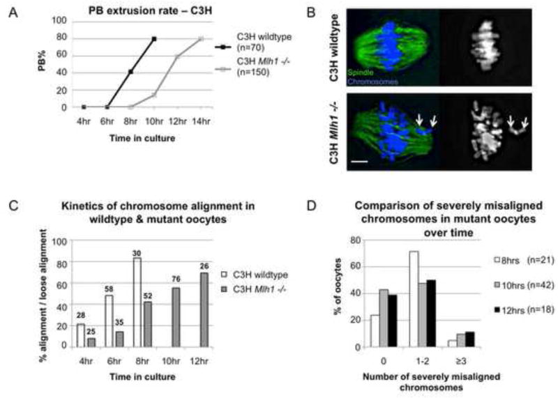
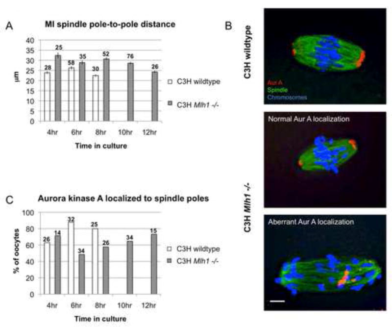
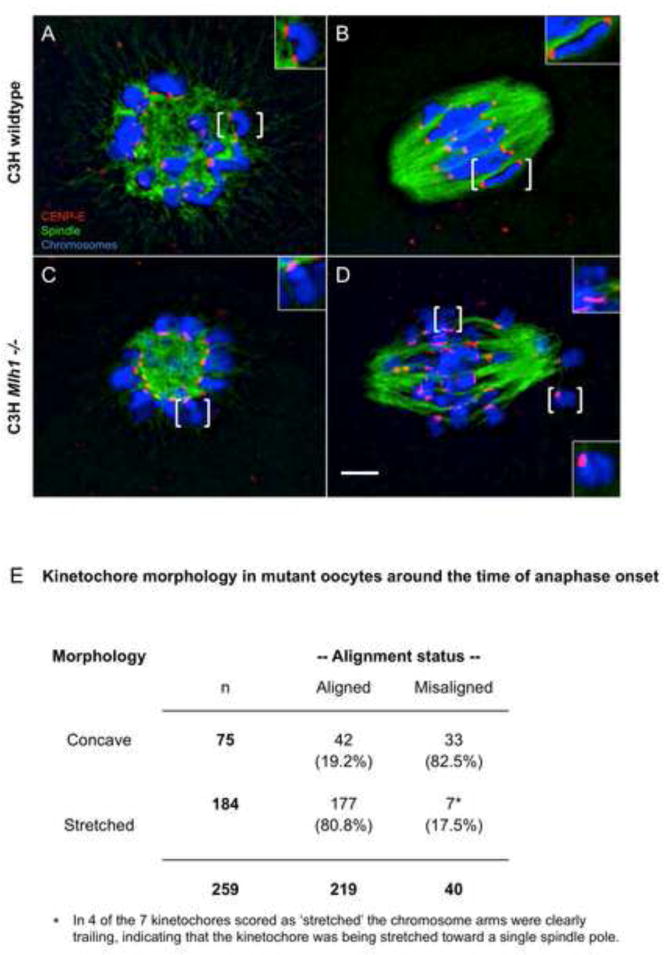
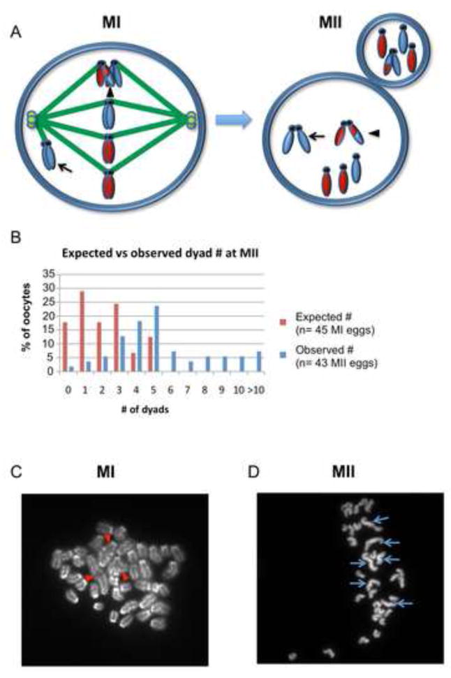
Segregation of homologs at the first meiotic division (MI) is facilitated by crossovers and by a physical constraint imposed on sister kinetochores that facilitates monopolar attachment to the MI spindle. Recombination failure or premature separation of homologs results in univalent chromosomes at MI, and univalents constrained to form monopolar attachments should be inherently unstable and trigger the spindle assembly checkpoint (SAC). Although univalents trigger cell-cycle arrest in the male, this is not the case in mammalian oocytes. Because the spindle assembly portion of the SAC appears to function normally, two hypotheses have been proposed to explain the lack of response to univalents: (1) reduced stringency of the oocyte SAC to aberrant chromosome behavior, and (2) the ability of univalents to satisfy the SAC by forming bipolar attachments. The present study of Mlh1 mutant mice demonstrates that metaphase alignment is not a prerequisite for anaphase onset and provides strong evidence that MI spindle stabilization and anaphase onset require stable bipolar attachment of a critical mass--but not all--of chromosomes. We postulate that subtle differences in SAC-mediated control make the human oocyte inherently error prone and contribute to the age-related increase in aneuploidy.


Figures

Figure 1
Figure 1. Oocytes from Mlh1 mutant females complete MI although a normal metaphase is not achieved
(A) In contrast to the meiotic arrest phenotype reported for Mlh1 mutant females on the B6 background ([17] and see Figure S4), prophase arrested oocytes meiotically matured in vitro from C3H mutant females extruded a polar body (PB) at wildtype frequency. However, by comparison with wildtype siblings, polar body extrusion was delayed by 3–4 hours in mutants. (B–D) To assess chromosome alignment, groups of oocytes were fixed at successive stages of meiotic maturation (i.e., after 4, 6, 8, 10, and 12 hours in culture), immunostained with an antibody to α-tubulin (green) to detect the spindle, counterstained with DAPI (blue) to visualize the chromosomes, imaged and scored for chromosome alignment. Because a normal metaphase configuration was not observed in mutant oocytes, cells were scored as “aligned” if more than 80% of chromosomes were loosely aligned at the spindle equator. (B) Representative images of cells scored as “aligned” and “loosely aligned.” Left images show spindle and chromosomes, right images show chromosomes alone. Top panels: Wildtype oocyte showing tight metaphase alignment of all chromosomes. Bottom panels: Mutant oocyte showing loose alignment of most chromosomes, but severe misalignment of two univalents (arrows). Scale bar = 5μm. (C) An analysis of the percentage of cells exhibiting aligned (wildtype) or loosely aligned (mutant) chromosomes after 4, 6, 8, 10, and 12 hours in culture demonstrates a delay in chromosome alignment in mutant oocytes. Numbers above each bar represent sample sizes. Note that mutants on the B6 background exhibited no improvement in chromosome alignment over time (Figure S4B). (D) Although, on average, 3–5 chromosomes were misaligned in cells scored as loosely aligned, an analysis of the number of severely misaligned chromosomes (those at or behind the poles) revealed no significant difference in cells collected after 8, 10, or 12 hours in culture (χ2 = 3.74, p=0.44).
Figure 2
Figure 2. MI spindle stabilization is delayed by aberrant chromosome behavior
(A) A comparison of MI spindle length demonstrates significant changes over time in both controls (F=7.06; p<0.001) and mutants (F=8.65; p<0.001), with the shortest spindle lengths occurring around the time of anaphase onset (8 hrs in controls; 12 hrs in mutants). Although spindle length was consistently greater in mutants, at the time of anaphase onset, lengths were not significantly different between controls and mutants (t=1.98; p>0.05). Error bars denote s.e.m. and numbers above each bar represent sample sizes. (B) Examples of Aurora kinase A localization during metaphase in a wildtype oocyte (top panel) and in Mlh1 mutant oocytes with normal Aurora kinase A localization (middle panel) and aberrant localization (bottom panel). Oocytes were immunostained with antibodies to α-tubulin (green) and Aurora kinase A (red) and counterstained with DAPI (blue). Scale bar = 5μm. (C) The kinetics of Aurora kinase A localization to the spindle poles in mutants and controls. Numbers above each bar represent sample sizes. Note that in mutants on the B6 background, aberrant Aurora kinase A localization persisted. (Figure S4C, D)
Figure 3
Figure 3. Kinetochore morphology is correlated with univalent behavior
Oocytes fixed at early prometaphase (A,C) and around the time of anaphase onset (8hrs in controls; 12 hrs in mutants) (B,D) from control and mutant females were immunostained with antibodies to α-tubulin (green) and CENP-E (red) to detect the spindle and kinetochores, respectively, and counterstained with DAPI (blue) to visualize the chromosomes. Brackets denote chromosomes shown in enlarged inset images. (A,C) In oocytes fixed during early prometaphase (i.e., after 4 hours in culture), the kinetochores of bivalents in wildtype oocytes (A) and of univalents in mutant oocytes (C) exhibited a single concave-shaped domain of CENP-E signal. (B) This morphology persisted in wildtype oocytes when kinetochores come under tension around the time of anaphase onset. (D) In mutant oocytes fixed around the time of anaphase onset, univalents aligned at the spindle equator typically exhibited a thin, stretched CENP-E signal. Scale bar = 5μm. Note that this morphology was not observed in mutant oocytes on the B6 background (Figure S4). (E) An analysis of 259 kinetochores on univalent chromosomes from 9 mutant oocytes collected around the time of anaphase onset demonstrates a highly significant correlation between alignment status and CENP-E morphology (χ2=65.9, p<0.001).
Figure 4
Figure 4. MII chromosome analysis provides evidence of intact segregation of univalents
(A) Schematic showing the segregation patterns that give rise to dyads and monads at MII. Homologs with an exchange (arrowhead, MI cell) will segregate reductionally, resulting in a dyad with genetically distinct chromatids (arrowhead, MII cell). In addition, a univalent that forms a monopolar attachment (arrow, MI cell) will segregate intact, resulting in a dyad with genetically identical sister chromatids (arrow, MII cell). Univalents that form bipolar attachments and segregate equationally will result in single chromatids (monads) at MII. (B) Comparison of the expected number of dyads with the number observed; expected values were based on data from the cytogenetic analysis of 45 MI oocytes (Figure S1) and assuming that dyads derive only from MI bivalents. (C) Representative image of an MI chromosome preparation showing 3 bivalents (red arrowheads) and 34 univalents. (D) Representative image of an MII chromosome preparation with 6 dyads (blue arrows) and 27 monads.

Comment in

References

    1. Musacchio A, Salmon ED. The spindle-assembly checkpoint in space and time. Nat Rev Mol Cell Biol. 2007;8:379–393. - PubMed
    1. Eaker S, Cobb J, Pyle A, Handel MA. Meiotic prophase abnormalities and metaphase cell death in MLH1-deficient mouse spermatocytes: insights into regulation of spermatogenic progress. Dev Biol. 2002;249:85–95. - PubMed
    1. Lipkin SM, Moens PB, Wang V, Lenzi M, Shanmugarajah D, Gilgeous A, Thomas J, Cheng J, Touchman JW, Green ED, et al. Meiotic arrest and aneuploidy in MLH3-deficient mice. Nat Genet. 2002;31:385–390. - PubMed
    1. Ward JO, Reinholdt LG, Motley WW, Niswander LM, Deacon DC, Griffin LB, Langlais KK, Backus VL, Schimenti KJ, O’Brien MJ, et al. Mutation in mouse hei10, an e3 ubiquitin ligase, disrupts meiotic crossing over. PLoS Genet. 2007;3:e139. - PMC - PubMed
    1. Burgoyne PS, Mahadevaiah SK, Turner JM. The consequences of asynapsis for mammalian meiosis. Nat Rev Genet. 2009;10:207–216. - PubMed

Publication types

MeSH terms