. 2011 Apr 26;21(8):681-6.
doi: 10.1016/j.cub.2011.03.030. Epub 2011 Apr 14.

Caveolin-1 deficiency causes cholesterol-dependent mitochondrial dysfunction and apoptotic susceptibility

Affiliations

Caveolin-1 deficiency causes cholesterol-dependent mitochondrial dysfunction and apoptotic susceptibility

Marta Bosch et al. Curr Biol. .

Abstract

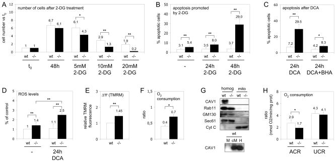
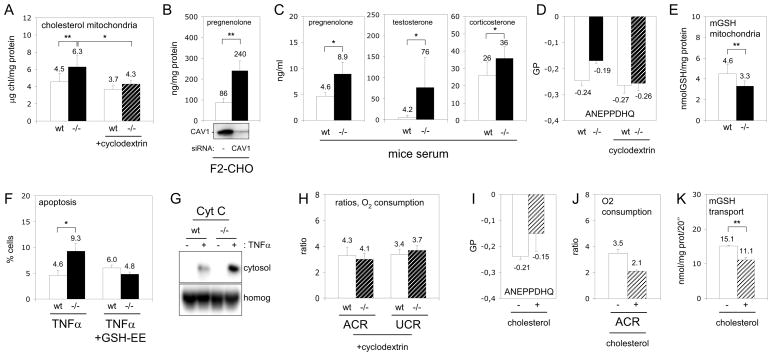
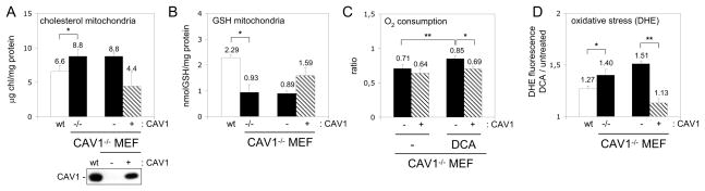
Caveolins (CAVs) are essential components of caveolae, plasma membrane invaginations with reduced fluidity, reflecting cholesterol accumulation. CAV proteins bind cholesterol, and CAV's ability to move between cellular compartments helps control intracellular cholesterol fluxes. In humans, CAV1 mutations result in lipodystrophy, cell transformation, and cancer. CAV1 gene-disrupted mice exhibit cardiovascular diseases, diabetes, cancer, atherosclerosis, and pulmonary fibrosis. The mechanism or mechanisms underlying these disparate effects are unknown, but our past work suggested that CAV1 deficiency might alter metabolism: CAV1(-/-) mice exhibit impaired liver regeneration unless supplemented with glucose, suggesting systemic inefficiencies requiring additional metabolic intermediates. Establishing a functional link between CAV1 and metabolism would provide a unifying theme to explain these myriad pathologies. Here we demonstrate that impaired proliferation and low survival with glucose restriction is a shortcoming of CAV1-deficient cells caused by impaired mitochondrial function. Without CAV1, free cholesterol accumulates in mitochondrial membranes, increasing membrane condensation and reducing efficiency of the respiratory chain and intrinsic antioxidant defense. Upon activation of oxidative phosphorylation, this promotes accumulation of reactive oxygen species, resulting in cell death. We confirm that this mitochondrial dysfunction predisposes CAV1-deficient animals to mitochondrial-related diseases such as steatohepatitis and neurodegeneration.


Figures

Figure 1
Figure 1. Mitochondrial dysfunction in CAV1−/− cells
(A) Wt (white bars) and CAV1−/− MEF (black bars) were cultured with 2-DG. After 48 hours cell number was determined and expressed with respect to the initial number of cells (t0). (B) Apoptosis, analysed by flow cytometry via binding of annexin V and staining with propidium iodide, promoted by 5mM 2-DG. (C) Apoptosis promoted by DCA, some cells were pre-treated with the antioxidant BHA. (D) Levels of ROS in cells incubated during 24 hours with DCA. The results are expressed as the relative H2DCFDA fluorescence with respect to untreated wt cells. (E) ΔΨ m of CAV1−/− with respect to wt MEF. (F) Oxygen consumption by wt and CAV1−/− MEF expressed as the routine flux control ratio. (G) Western blotting analysis of CAV1 (plasma membrane), Rab11 (recycling endosomes), GM130 (Golgi complex), Sec61 (ER) and cytochrome C (Cyt C, mitochondria) in purified wt and CAV1−/− mitochondria (M), homogenates (H) and in a crude fraction that contains mitochondria and associated ER (cM). (H) Ratios of oxygen consumption in wt (white bars) and CAV1−/− mitochondria (black bars) purified from mice liver. Statistical significances were determined in at least 5 independent experiments or 10 mice using the Student’s t test, *P<0.05, **P<0.01.
Figure 2
Figure 2. Cholesterol accumulation promotes dysfunction of CAV1−/− mitochondria
(A) Free cholesterol in wt (white bars) and CAV1−/− (black bars) mitochondria purified from mice liver. In some experiments mitochondria were pre-treated with cyclodextrin to extract cholesterol (slashed bars). (B) Expression of CAV1 in F2-CHO cells was reduced by RNA interference during 48 hours (Western blotting of CAV1 is shown in the bottom) and production of pregnenolone was measured during the next 24 hours. (C) Pregnenolone, corticosterone and testosterone levels in the serum of CAV1−/− (black bars) and wt mice (white bars). (D) Membrane order analysed with ANEPPDHQ of wt (white bars), CAV1−/− (black bars) and cyclodextrin-treated wt white bars) and CAV1−/− purified mitochondria (slashed bars). (E) Mitochondrial GSH in wt (white bars) and CAV1−/− (black bars) mitochondria purified from mice liver. (F) Apoptosis promoted by 24 hours of TNFα in untreated wt (white bars) and CAV1−/− MEF (black bars) or in cells treated with GSH-EE. (G) Cytochrome C (Cyt C) in cytosolic supernatants and homogenates (homog) corresponding to TNFα treated MEFs. (H) Purified mitochondria from wt (with bars) and CAV1−/− (slashed bars) were treated with cyclodextrin and the rates of oxygen consumption measured. (I–J) Purified wt mitochondria (white bars) were enriched with 25% of cholesterol (slashed bars) and membrane condensation (I) and rates of oxygen consumption (J) were measured. (K) Influx of a radio-labelled GSH into wt mitochondria untreated (white bars) or enriched with 25% of cholesterol (slashed bars).
Figure 3
Figure 3. Re-expression of CAV1 recovers mitochondrial function
(A and B) Free cholesterol and GSH in mitochondria purified from wt (white bars), CAV1−/− (black bars), CAV1−/−-reconstituted MEFs (slashed bars) and CAV1−/− MEF infected with an empty vector (black bars). CAV1 levels are shown by Western blotting. (C) Routine flux control ratio in untreated MEFs and in cells incubated with DCA for 5 hours. (D) Oxidative stress caused by mitochondrial function in MEFs. Results are expressed as the ratio between the fluorescence intensity of DHE after treating the cells with DCA for 5 hours with respect to the initial intensity.
Figure 4
Figure 4. Dysfunction of CAV1−/− mitochondria enhances pathogenesis
(A–C) To model steatohepatitis wt (white bars) and CAV1−/− mice (black bars) were treated with Jo2. Liver damage was evaluated 24 hours later by appearance of transaminases in serum (AST and ALT). Inflammation was visualised in liver sections of wt (left) and CAV1−/− mice (right) with hematoxiline/eosin and myeloperoxidase staining. (D–E) Wt and CAV1−/− primary hepatocytes were treated with Jo2 for 24 hours. Apoptosis in wt (left panel) and CAV1−/− hepatocytes (right) was visualized with a Hoechst staining (D) and released cytochrome C (CytC) into the cytosol quantified by Western blot (E). (F) MTT cell viability assay of wt and CAV1−/− hepatocytes treated with Jo2 or with Jo2/GSH-EE. (G–H) Free cholesterol and mGSH in wt (white bars) and CAV1−/− (black bars) purified brain mitochondria. (I) ROS generation in wt (white bars) and CAV1−/− (black bars) purified brain mitochondria (some treated with cyclodextrin, slashed bars) incubated with Aβ1–42. (J–K) 3-NP was injected in the striatum of wt and CAV1−/− mice and the volume of the lesion measured 24 hours later in serial Fluoro-Jade-stained sections and apoptotic nucleus were visualised in TUNEL-stained sections (K) of wt (left) and CAV1−/− striatum (right).

References

    1. Parton RG, Simons K. The multiple faces of caveolae. Nat Rev Mol Cell Biol. 2007;8:185–194. - PubMed
    1. Pol A, Luetterforst R, Lindsay M, Heino S, Ikonen E, Parton RG. A caveolin dominant negative mutant associates with lipid bodies and induces intracellular cholesterol imbalance. J Cell Biol. 2001;152:1057–1070. - PMC - PubMed
    1. Pol A, Martin S, Fernandez MA, Ingelmo-Torres M, Ferguson C, Enrich C, Parton RG. Cholesterol and fatty acids regulate dynamic caveolin trafficking through the Golgi complex and between the cell surface and lipid bodies. Mol Biol Cell. 2005;16:2091–2105. - PMC - PubMed
    1. Cao H, Alston L, Ruschman J, Hegele RA. Heterozygous CAV1 frameshift mutations (MIM 601047) in patients with atypical partial lipodystrophy and hypertriglyceridemia. Lipids Health Dis. 2008;7:3. - PMC - PubMed
    1. Kim CA, Delepine M, Boutet E, El Mourabit H, Le Lay S, Meier M, Nemani M, Bridel E, Leite CC, Bertola DR, et al. Association of a homozygous nonsense caveolin-1 mutation with Berardinelli-Seip congenital lipodystrophy. J Clin Endocrinol Metab. 2008;93:1129–1134. - PubMed

Publication types