. 2011 Apr 22;34(4):492-504.
doi: 10.1016/j.immuni.2011.03.017. Epub 2011 Apr 14.

Asymmetric proteasome segregation as a mechanism for unequal partitioning of the transcription factor T-bet during T lymphocyte division

Affiliations

Asymmetric proteasome segregation as a mechanism for unequal partitioning of the transcription factor T-bet during T lymphocyte division

John T Chang et al. Immunity. .

Abstract

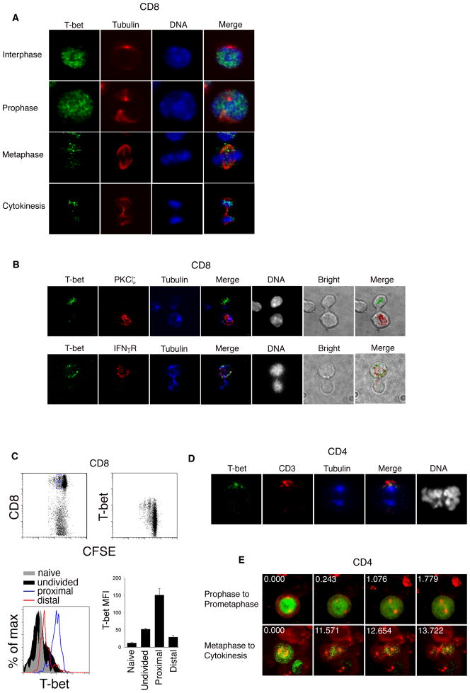
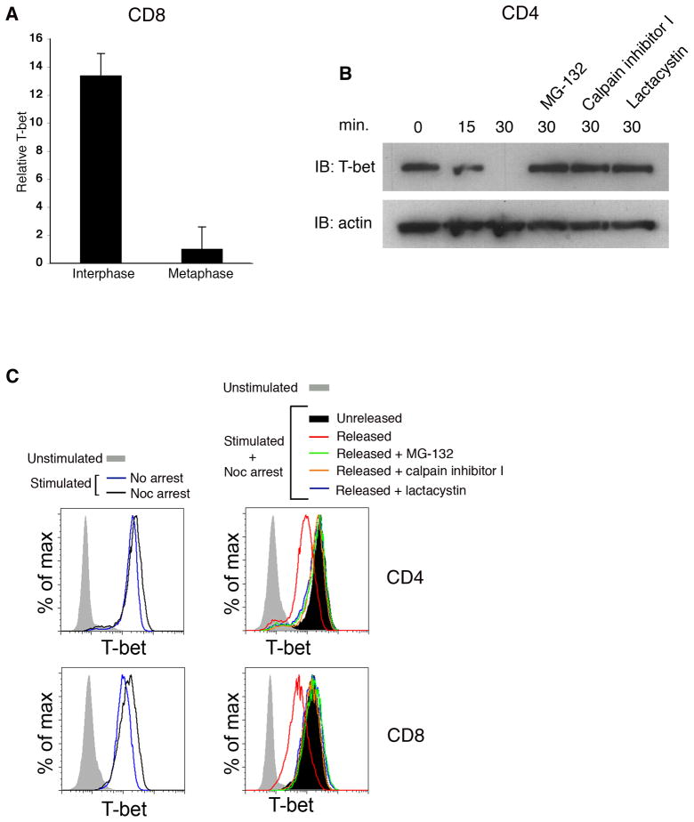
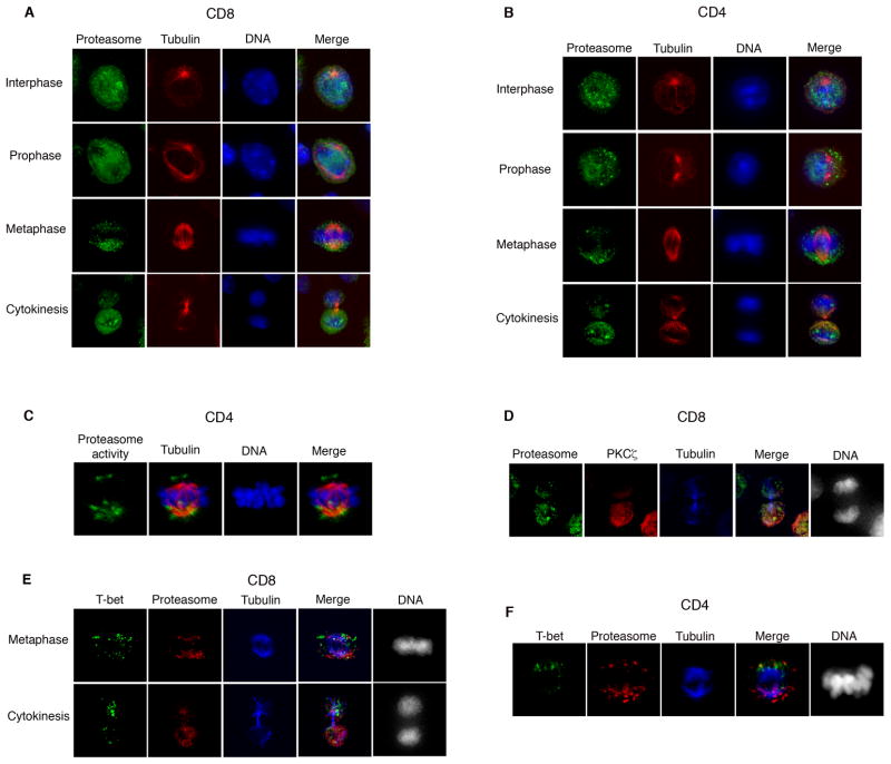
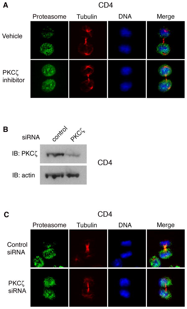
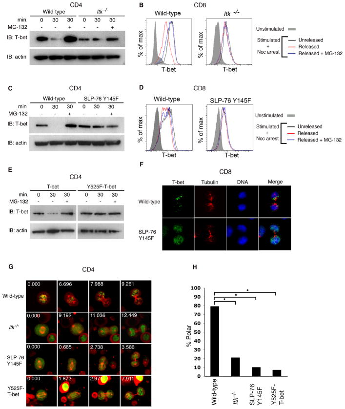
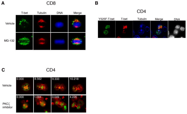
Polarized segregation of proteins in T cells is thought to play a role in diverse cellular functions including signal transduction, migration, and directed secretion of cytokines. Persistence of this polarization can result in asymmetric segregation of fate-determining proteins during cell division, which may enable a T cell to generate diverse progeny. Here, we provide evidence that a lineage-determining transcription factor, T-bet, underwent asymmetric organization in activated T cells preparing to divide and that it was unequally partitioned into the two daughter cells. This unequal acquisition of T-bet appeared to result from its asymmetric destruction during mitosis by virtue of concomitant asymmetric segregation of the proteasome. These results suggest a mechanism by which a cell may unequally localize cellular activities during division, thereby imparting disparity in the abundance of cell fate regulators in the daughter cells.


Figures

Figure 1
Figure 1. Asymmetric partitioning of T-bet in dividing T lymphocytes
(A) Undivided P14 CD8+ TCR transgenic T cells were adoptively transferred into wild-type recipients infected with gp33-L. monocytogenes, harvested at 36h after transfer, and examined by confocal microscopy after staining for T-bet (green), β-tubulin (red), and DNA (blue). Asymmetry of T-bet inheritance was observed in 66% of cells (n=80). (B) Undivided P14 CD8+ TCR transgenic T cells were harvested as in (A) and stained for T-bet (green), PKCζ or IFNγR (red), β-tubulin (blue), and DNA (grayscale). In co-staining experiments where both molecules were asymmetrically inherited, T-bet and the IFNγR were inherited by the same daughter cell in 100% (n=15) of cells, and T-bet and PKCζ were inherited by opposite daughters in 87% (n=14) of cells. (C) CFSE-labeled P14 transgenic CD8+ T cells from recipients infected with gp33-L. monocytogenes were harvested 48h after infection, stained with antibodies to detect CD8 and T-bet, and analyzed by flow cytometry. Putative proximal and distal daughter cells (detected as the second brightest CFSE peak) were gated on the basis of CD8 expression (gates shown in upper left panel). T-bet mean fluorescence intensity (MFI) of the gated proximal (blue histogram), distal (red histogram), naïve (gray filled histogram), and undivided (black filled histogram) populations is shown. Error bars indicate standard error of the mean (SEM). (D) CD4+ T cells were activated in vitro and stained for T-bet (green), CD3 (red), β-tubulin (blue), and DNA (grayscale). In co-staining experiments where both molecules were asymmetrically inherited, T-bet and CD3 were inherited by the same daughter cell in 100% (n=16) of cells. (E) CD4+ T cells were activated in vitro and transduced at 48h with T-bet-GFP and cherry-alpha-tubulin fusion proteins. After 3d, T cells were restimulated in vitro for 24h and imaged by time-lapse confocal microscopy. Prophase cells were imaged through prometaphase (upper panel), and metaphase cells imaged through cytokinesis (lower panel). Asymmetric T-bet partitioning by the daughter cells was observed in 68% of cells (n=23). Relative time (in minutes) is indicated in the upper left-hand corner of each panel. Results are representative of three separate experiments.
Figure 2
Figure 2. T-bet undergoes proteasome-dependent degradation during mitosis
(A) Quantification of T-bet signal in interphase vs. metaphase T cells represented in Figure 1A. T-bet signal was compared between interphase and metaphase blasts imaged in the same field of view (n=61). Error bars indicate SEM. (B) CD4+ T cells were activated in vitro for 24h and then synchronized with nocodazole. Cells were washed free of drug, cultured in vitro with or without the proteasome inhibitors MG-132, calpain inhibitor I, or lactacystin. Cell lysates were prepared at 0, 15, or 30m after nocodazole washout. T-bet and β-actin levels were assessed by immunoblotting. (C) CD4+ or CD8+ T cells were prepared as in (B) and T-bet levels assessed by intracellular staining at 0 and 30m after nocodazole washout. T-bet levels of naïve T cells and activated cells not treated with nocodazole (“freely cycling”) are also shown. Results are representative of three separate experiments.
Figure 3
Figure 3. Unequal proteasomal segregation as a mechanism for asymmetric cell division
(A)Undivided P14 CD8+ TCR transgenic T cells were harvested as in Figure 1A and stained for the α1 chain of the proteasome 20S subunit (green), β-tubulin (red), and DNA (blue). Asymmetry of proteasome localization was observed in 62% (n=74) of cells. (B) CD4+ T cells were activated in vitro for 28h and stained as in (A). Asymmetry of proteasome localization was observed in 74% (n=125) of cells. (C) CD4+ T cells were activated in vitro for 28h, treated with the proteasome activity probe MVB003 for 2h, and stained for β-tubulin (red), and DNA (blue). Asymmetry of degradative activity was observed in 65% (n=22) of cells. (D) Undivided P14 CD8+ TCR transgenic T cells were harvested as in Figure 1A, and stained for the proteasome 20S α1 subunit (green), PKCζ (red), β-tubulin (blue), and DNA (grayscale). In co-staining experiments where both molecules were asymmetrically inherited, proteasome and PKCζ were inherited by the same daughter cell in 95% of cells (n=34). (E) Undivided P14 CD8+ TCR transgenic T cells were harvested as in Figure 1A, and stained for T-bet (green), proteasome 20S α1 subunit (red), β-tubulin (blue), and DNA (grayscale). In co-staining experiments where both molecules were asymmetrically inherited, proteasome and T-bet were inherited by opposite daughter cells in 90% of cells (n=29). (F) CD4+ T cells were activated as in (B) and stained as in (E). In co-staining experiments where both molecules were asymmetrically inherited, proteasome and T-bet were inherited by opposite daughter cells in 90% of cells (n=14). Results are representative of three separate experiments.
Figure 4
Figure 4. Asymmetry of the proteasome may depend on the polarity network
(A) CD4+ T cells were activated in vitro for 28h, treated for 1h with vehicle or a pharmacologic inhibitor of PKCζ, and stained for the proteasomal 20S α1 subunit (green), β-tubulin (red), and DNA (blue). Asymmetry of proteasome localization was observed in 61% (n=25) of vehicle-treated vs. 27% (n=29) of PKCζ inhibitor treated cells (p<0.001). (B) CD4+ T cells were activated in vitro for 48h, and control or PKCζ siRNA was introduced using electroporation. Lysates were prepared 72h later, and PKCζ and β-actin levels assessed by immunoblotting. (C) CD4+ T cells were transfected with control or PKCζ siRNA as in (B). 48h later, cells were restimulated in vitro for 24h and stained with the proteasomal 20S α1 subunit (green), β-tubulin (red), and DNA (blue). Asymmetry of proteasome localization was observed in 63% (n=60) of control transfected vs. 32% (n=62) of PKCζ siRNA transfected cells (p<0.001). Results are representative of two separate experiments.
Figure 5
Figure 5. Mutations preventing phosphorylation of T-bet impair its proteasome-dependent degradation and asymmetric partitioning during mitosis
(A) CD4+ T cells from wild-type or Itk−/− mice were activated in vitro and synchronized with nocodazole as in Figure 2B. After drug washout, cells were cultured with or without MG-132 for 30m. Cell lysates were prepared at 0 or 30m after drug washout. T-bet and β-actin levels were assessed by immunoblotting. (B) CD8+ T cells from wild-type or Itk−/− mice were activated as in (A) and T-bet levels assessed by intracellular staining at 0 or 30m after drug washout. (C) CD4+ T cells from wild-type and SLP-76 Y145F mice were activated and analyzed as in (A). (D) CD8+ T cells from wild-type and SLP-76 Y145F mice were activated and analyzed as in (B). (E) CD4+ T cells from T-bet-deficient mice were transduced with retroviruses encoding wild-type T-bet-GFP or Y525F-T-bet-GFP. After 3d, cells were restimulated for 24h and synchronized with nocodazole and analyzed as in (A). (F) Wild-type or SLP-76 Y145F P14 CD8+ TCR transgenic T cells were adoptively transferred into wild-type recipients infected with gp33-L. monocytogenes, harvested at 36h after transfer, and stained for T-bet (green), β-tubulin (red), and DNA (blue). Asymmetric partitioning of T-bet was observed in 72% (n=21) of wild-type versus 15% (n=26) of SLP-76 Y145F P14 CD8+ T cells (p<0.001). (G) CD4+ T cells from wild-type, Itk−/−, and SLP-76 Y145F mice were transduced with cherry-alpha-tubulin and either T-bet-GFP or Y525F-T-bet-GFP. Cells were imaged as in Figure 1E. (H) Quantification of asymmetric T cell partitioning into daughter cells represented in (G). The number of cells transduced with T-bet-GFP that were examined in each group: wild-type (46), Itk−/− (48), SLP-76 Y145F (29). The number of wild-type cells transduced with Y525F-T-bet-GFP examined was 27. p<0.001 (*). Results are representative of two separate experiments.
Figure 6
Figure 6. Preventing degradation of T-bet disrupts the asymmetric partitioning of T-bet
(A) Undivided P14 CD8+ TCR transgenic T cells were harvested as in Figure 1A and cultured in vitro with vehicle or MG-132 for 4h prior to staining with T-bet (green), β-tubulin (red), and DNA (blue). (B) CD4+ T cells from T-bet-null mice were transduced with both wild-type T-bet-cherry and Y525F-T-bet-GFP, restimulated in vitro and stained for β-tubulin (blue). Cytokinetic cells expressing both wild-type T-bet (red) and Y525F-Tbet (green) were scored. Asymmetry of wild-type T-bet and Y525F-T-bet was observed in 60% and 9% (n=23) of cells, respectively (p<0.001). (C) CD4+ T cells were transduced as in Figure 1E. After 3d, cells were restimulated for 24h, treated for 1h with vehicle (top panel) or PKCζ inhibitor (bottom panel), and imaged as in Figure 1E. Asymmetric partitioning of T-bet occurred in 82% (n=28) of vehicle-treated versus 14% (n=42) of PKCζ inhibitor-treated cells (p<0.001). Results are representative of two separate experiments.

Comment in

Similar articles

Cited by

References

    1. Afkarian M, Sedy JR, Yang J, Jacobson NG, Cereb N, Yang SY, Murphy TL, Murphy KM. T-bet is a STAT1-induced regulator of IL-12R expression in naive CD4+ T cells. Nat Immunol. 2002;3:549–557. - PubMed
    1. Atherly LO, Lucas JA, Felices M, Yin CC, Reiner SL, Berg LJ. The Tec family tyrosine kinases Itk and Rlk regulate the development of conventional CD8+ T cells. Immunity. 2006;25:79–91. - PubMed
    1. Azar GA, Lemaitre F, Robey EA, Bousso P. Subcellular dynamics of T cell immunological synapses and kinapses in lymph nodes. Proc Natl Acad Sci U S A. 2010;107:3675–3680. - PMC - PubMed
    1. Barcia C, Thomas CE, Curtin JF, King GD, Wawrowsky K, Candolfi M, Xiong WD, Liu C, Kroeger K, Boyer O, et al. In vivo mature immunological synapses forming SMACs mediate clearance of virally infected astrocytes from the brain. J Exp Med. 2006;203:2095–2107. - PMC - PubMed
    1. Berg LJ. Signalling through TEC kinases regulates conventional versus innate CD8(+) T-cell development. Nat Rev Immunol. 2007;7:479–485. - PubMed

Publication types

Substances