Review
. 2011 Jul;193(4):354-61.
doi: 10.1016/j.aanat.2011.02.013. Epub 2011 Mar 12.

Enhancing recovery from peripheral nerve injury using treadmill training

Affiliations
Review

Enhancing recovery from peripheral nerve injury using treadmill training

Arthur W English et al. Ann Anat. 2011 Jul.

Abstract

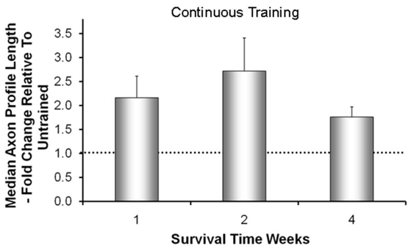
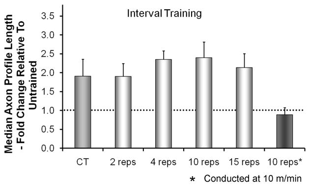
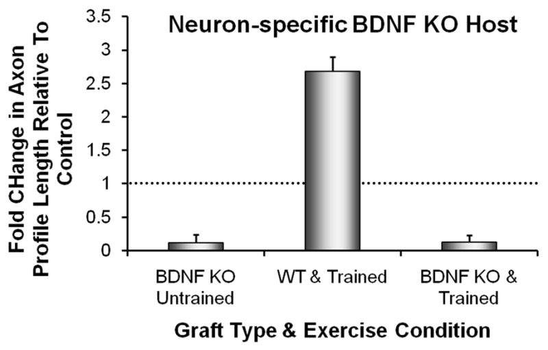
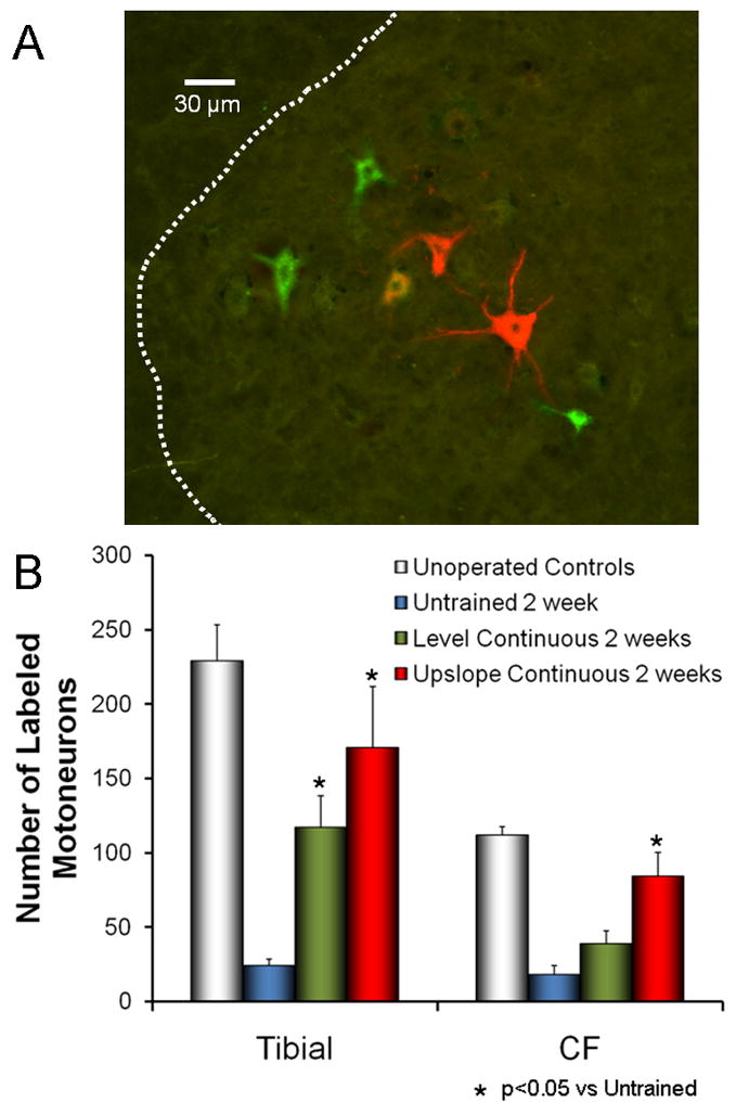
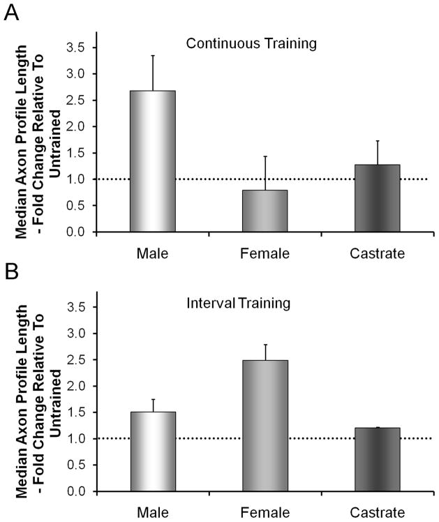
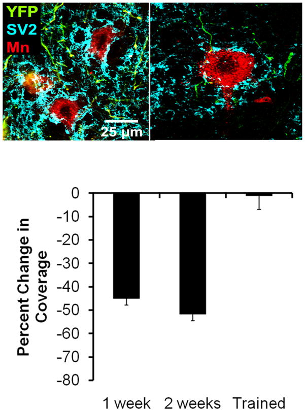
Full functional recovery after traumatic peripheral nerve injury is rare. We postulate three reasons for the poor functional outcome measures observed. Axon regeneration is slow and not all axons participate. Significant misdirection of regenerating axons to reinnervate inappropriate targets occurs. Seemingly permanent changes in neural circuitry in the central nervous system are found to accompany axotomy of peripheral axons. Exercise in the form of modest daily treadmill training impacts all three of these areas. Compared to untrained controls, regenerating axons elongate considerably farther in treadmill trained animals and do so via an autocrine/paracrine neurotrophin signaling pathway. This enhancement of axon regeneration takes place without an increase in the amount of misdirection of regenerating axons found without training. The enhancement also occurs in a sex-dependent manner. Slow continuous training is effective only in males, while more intense interval training is effective only in females. In treadmill trained, but not untrained mice the extent of coverage of axotomized motoneurons is maintained, thus preserving important elements of the spinal circuitry.


Figures

Figure 1
Figure 1
The effects of Continuous treadmill training on axon regeneration in peripheral nerves. Cut common fibular nerves in thy-1-YFP-H mice were repaired with a short length of nerve harvested from a non-fluorescent littermate. Animals were trained continuously for one hour per day at a slow treadmill speed (10 m/min), five days per week for no more than two weeks. Using images of optical sections made through these grafts, the lengths of YFP+ regenerating axon profiles were measured at different survival times. Average median axon profile lengths, expressed as a percentage of untrained controls (±SEM, N=4 for each), are shown. The horizontal dashed line at 1.0 indicates the length of regenerating axons in untrained controls. Data are from Sabatier et al (2008).
Figure 2
Figure 2
The effects of Interval treadmill training on axon regeneration in peripheral nerves. The paradigm used was similar to that described for Figure 1 except for the pattern of treadmill training. Animals were trained at a faster treadmill speed (20 m/min), for two minutes and then rested for five minutes. This interval was repeated different numbers of times. Training was conducted five days/week for two weeks. Average median axon profile lengths, expressed as a percentage of untrained controls (±SEM, N=4 for each), are shown. The horizontal dashed line at 1.0 indicates the length of regenerating axons in untrained controls. Data are from Sabatier et al (2008).
Figure 3
Figure 3
Axon regeneration is not enhanced by treadmill training in neuron-specific BDNF knockout mice. Cut common fibular and tibial nerves in female SLICK::BDNFf/f host mice were repaired with a short length of nerve harvested from non-fluorescent, strain-matched graft donor mice. Some graft donor mice were wild type (WT) and others were systemic BDNF knockout mice (BDNF KO). In the host mice in these experiments, the gene for BDNF was knocked out in motoneurons expressing YFP. The cut YFP+ axons in these mice were constrained to regenerate into an environment lacking (BDNF KO) or containing (WT) BDNF. Animals were trained using an interval training paradigm and the lengths of profiles of YFP+ regenerating axons measured in grafts after two weeks were compared to those found in untrained WT mice whose axons regenerated into WT grafts (Controls). Average median axon profile lengths, expressed as a percentage of untrained Controls (+SEM, N=4 for each), are shown from measurements made in grafts from BDNF KO mice (Untrained on the left, treadmill Trained on the right) and in WT mice (center). The horizontal dashed line at 1.0 indicates the length of regenerating axons in untrained Controls. Data are from Wilhelm et al (2009).
Figure 4
Figure 4
The effects of upslope training on axon regeneration in cut peripheral nerves. The sciatic nerves of mice were cut and repaired by end-to-end anastomosis. Some mice were trained using a continuous training paradigm with the treadmill level and others with the treadmill inclined upwards at 20 degrees. Two weeks after transection, the tibial and common fibular branches were cut 4 mm distal to the original transection and exposed to different retrograde fluorescent tracers. A. Examples of motoneurons retrogradely labeled from red and green fluorescent tracers applied to the tibial and common fibular nerves, respectively. B. Labeled motoneurons with axons regenerating that distance into these two branches were counted in histological sections of the lumbar spinal cord. Comparisons were made between the two groups of trained mice and to intact and untrained (both lesioned and unlesioned) mice. Each bar represents the mean number of labeled motoneurons (±SEM, N=4 for each).
Figure 5
Figure 5
Sex differences in the effects of treadmill training on axon regeneration in cut peripheral nerves. In each graph, average median axon profile lengths, expressed as a percentage of untrained controls (±SEM, N=4 for each), are shown. The horizontal dashed line at 1.0 indicates the length of regenerating axons in untrained controls. A. If mice are trained using a continuous training paradigm, enhancement is found only in males and not females or castrated males. B. If mice are exercised at intervals, enhanced regeneration is found only in females, not in males or castrated males.
Figure 6
Figure 6
Effects of treadmill training on synaptic stripping following peripheral nerve transection. Images of labeled motoneurons in untrained intact mice and one week following sciatic nerve transection are shown in the top two panels. In these mice, a subset of neurons express yellow fluorescent protein (YFP, shown in green in these panels) and motoneurons innervating the gastrocnemius muscle were marked by injection of a red fluorescent retrograde tracer (cholera toxin B-Alexafluor 555) four days prior to sciatic nerve transection (without repair). Expression of the ubiquitous synaptic vesicle protein, SV2, was demonstrated using immuno-histofluorescence, using a secondary antibody conjugated to Alexafluor 647 (shown as cyan in these panels). Note the robust SV2 immunoreactivity in the intact mouse and the relative paucity of synaptic coverage one week after nerve transection. In a series of trained and untrained mice, the proportion of the soma and proximal dendrites of retrogradely labeled motoneurons contacted by SV2 immune-positive structures was determined. The average change in this synaptic coverage was determined by comparison to coverage in intact and untrained mice and is shown in the bottom panel. Each bar represents the average percent change in synaptic coverage (±SEM, N=4 for each) in untrained mice after 1 or 2 weeks and mice treadmill trained for two weeks. Note that the reduction in coverage of nearly 50% observed in untrained mice is completely absent in trained mice.

Similar articles

Cited by

References

    1. Adlard PA, Cotman CW. Voluntary exercise protects against stress-induced decreases in brain-derived neurotrophic factor protein expression. Neuroscience. 2004;124:985–992. - PubMed
    1. Adlard PA, Perreau VM, Engesser-Cesar C, Cotman CW. The timecourse of induction of brain-derived neurotrophic factor mRNA and protein in the rat hippocampus following voluntary exercise. Neurosci Lett. 2004;363:43–48. - PubMed
    1. Alvarez FJ, Bullinger KL, Titus HE, Nardelli P, Cope TC. Permanent reorganization of Ia afferent synapses on motoneurons after peripheral nerve injury. Ann NY Acad Sci. 2010;1198:231–241. - PMC - PubMed
    1. American Physiological Society. Resource Book for the Design of Animal Exercise Protocols. American Physiological Society; 2006.
    1. Asensio-Pinilla E, Udina E, Jaramillo J, Navarro X. Electrical stimulation combined with exercise increase axonal regeneration after peripheral nerve injury. Exp Neurol. 2009;219:258–265. - PubMed

Publication types

Substances