. 2011 Jun 1;17(11):3512-9.
doi: 10.1158/1078-0432.CCR-09-2358. Epub 2011 Apr 15.

New strategies in Barrett's esophagus: integrating clonal evolutionary theory with clinical management

Affiliations

New strategies in Barrett's esophagus: integrating clonal evolutionary theory with clinical management

Brian J Reid et al. Clin Cancer Res. .

Abstract

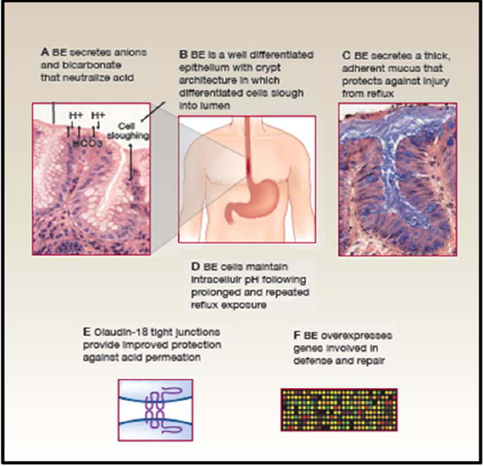
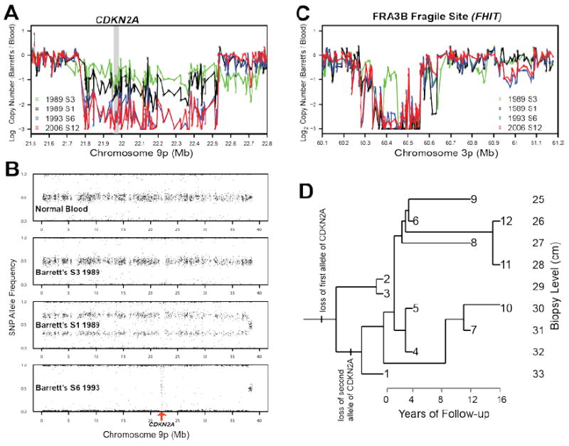
Barrett's esophagus is a condition in which the normal stratified squamous epithelium of the distal esophagus is replaced by intestinal metaplasia. For more than three decades, the prevailing clinical paradigm has been that Barrett's esophagus is a complication of symptomatic reflux disease that predisposes to esophageal adenocarcinoma. However, no clinical strategy for cancer prevention or early detection based on this paradigm has been proven to reduce esophageal adenocarcinoma mortality in a randomized clinical trial in part because only about 5% to 10% of individuals with Barrett's esophagus develop esophageal adenocarcinoma. Recent research indicates that Barrett's metaplasia is an adaptation for mucosal defense in response to chronic reflux in most individuals. The risk of progressing to esophageal adenocarcinoma is determined by development of genomic instability and dynamic clonal evolution in the distal esophagus modulated by host and environmental risk and protective factors, including inherited genotype. The challenge for investigators of Barrett's esophagus lies in integrating knowledge about genomic instability and clonal evolution into clinical management to increase the lifespan and quality of life of individuals with this condition.


Figures

Figure 1
Figure 1. Barrett’s specialized intestinal metaplasia and mucosal defense
Barrett’s metaplasia arises in an environment of chronic reflux in which the distal esophagus is exposed to high levels of local and systemic damage from acid, bile, and tobacco products as well as the inflammatory responses to the injury(12, 55-60). All are mutagenic. Barrett’s metaplasia has a number of defenses against this mutagenic environment that are not found in esophageal squamous epithelium(12, 43). A. Barrett’s metaplasia secretes anions, including bicarbonate, that participate in buffering acid reflux (61). B. Barrett’s metaplasia is a well differentiated epithelium with crypt architecture in which putative stem cells residing at the base give rise to proliferating transient amplifying cells and differentiated cells that are sloughed into the lumen. This architecture has been proposed to be tumor suppressive because mutations in transient amplifying or differentiated non-stem cells would be shed from the body before they could accumulate the serial mutations that lead to cancer(62). C Barrett’s metaplasia secretes a thick adherent mucus not present in squamous esophageal epithelium for defense against acid and bile reflux(45, 46, 63)(64). D. Barrett’s esophageal cells maintain physiological intracellular pH following prolonged and repeated reflux exposure(65). E. The tight junctions of Barrett’s metaplasia overexpress claudin 18 and several other claudins, including 1, 4, 12 and 23, that provide protection against acid permeation(66). F. A combined expression and proteomics study of Barrett’s metaplasia reported overexpression of genes involved in mucosal defense and repair(44).
Figure 2
Figure 2. Benign clonal evolution in one patient with Barrett’s esophagus studied longitudinally over 16 years
Purified Barrett’s epithelium from endoscopic biopsies was assayed with Illumina 317K SNP arrays and compared to a blood sample control. (A) Copy number analysis, normalized by SNP intensities from blood, reveal a single copy loss at CDKN2A in samples 2 (data not shown) and 3 in 1989, but homozygous deletion in CDKN2A in sample 1 and all samples from following years. At first endoscopy, in 1989, two clones were detected, one with a small deletion of one allele at the CDKN2A locus, and the other with copy neutral LOH of the entire 9p arm with the CDKN2A deleted allele, generating biallelic deletion at CDKN2A. (B) The SNP allele frequencies reveal a focal deletion in the CDKN2A locus in samples 2 and 3 in 1989, but sample 1 included a mixture of the clone from samples 2 and 3 with a new clone with copy neutral LOH of 9p and biallelic deletion of CDKN2A. All samples from 1993 and later show that the clone with biallelic deletion of CDKN2A went to fixation, leading to random noise in the allele frequencies for the SNPs in that region, seen in the vertical (“waterfall”) band in the bottom panel of B. The fact that the rest of the 9p arm remains diploid can be seen in the copy number data (A). The clone with deletion of the single allele of CDKN2A, which extends past 22.5Mb on chromosome 9p, also had a single deletion in fragile site FRA3B at 60.42Mb that distinguished it from the other clones (C). This and other lesions of the clone in samples 2 and 3 were never observed again after 1989, suggesting that that clone was driven extinct by the clone from sample 1, with biallelic deletion of CDKN2A. A Camin-Sokal maximum parsimony reconstruction of the genealogy of clones (D), based on polymorphic copy number lesions in 283 loci across the entire genome in the Barrett’s biopsies, shows that there was only one large clonal expansion, between 1989 and 1993. After 1993, the Barrett’s segment remained stable, with the accumulation of small interstitial lesions but no clonal expansions, no aneuploidy and no progression to cancer.

References

    1. Wang KK, Sampliner RE. Updated guidelines 2008 for the diagnosis, surveillance and therapy of Barrett’s esophagus. Am J Gastroenterol. 2008;103:788–97. - PubMed
    1. Barrett N. Chronic peptic ulcer of the oesophagus and ‘oesophagitis’. Br J Surg. 1950;38:175–82. - PubMed
    1. Naef AP, Savary M, Ozzello L. Columnar-lined lower esophagus: an acquired lesion with malignant predisposition. Report on 140 cases of Barrett’s esophagus with 12 adenocarcinomas. Journal of Thoracic and Cardiovascular Surgery. 1975;70:826–35. - PubMed
    1. Sampliner RE. Practice guidelines on the diagnosis, surveillance, and therapy of Barrett’s esophagus. Am J Gastroenterol. 1998;93:1028–32. - PubMed
    1. Thomas T, Abrams KR, De Caestecker JS, Robinson RJ. Meta analysis: Cancer risk in Barrett’s oesophagus. Aliment Pharmacol Ther. 2007;26:1465–77. - PubMed

Publication types