

Review
doi: 10.1093/cvr/cvr116.
Epub 2011 Apr 15.
Pharyngeal mesoderm development during embryogenesis: implications for both heart and head myogenesis
Affiliations
- PMID: 21498416
- PMCID: PMC3125075
- DOI: 10.1093/cvr/cvr116
Item in Clipboard
Review
Pharyngeal mesoderm development during embryogenesis: implications for both heart and head myogenesis
Cardiovasc Res.
.
Abstract
The pharyngeal mesoderm (PM), located in the head region of the developing embryo, recently triggered renewed interest as the major source of cells contributing to broad regions of the heart as well as to the head musculature. What exactly is PM? In this review, we describe the anatomical and molecular characteristics of this mesodermal population and its relationship to the first and second heart fields in chick and mouse embryos. The regulatory network of transcription factors and signalling molecules that regulate PM development is also discussed. In addition, we summarize recent studies into the evolutionary origins of this tissue and its multipotential contributions to both cardiac and pharyngeal muscle progenitors.
Figures
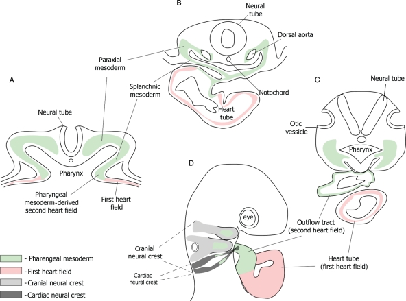
Pharyngeal mesoderm cells give rise to parts of the heart and the pharyngeal muscles. (A–D) Schematic illustration of the anatomy of the pharyngeal mesoderm in sections of a 1.5–3-day-old chick embryo. Pharyngeal mesoderm cells (green) in the anterior part of the embryo surround the pharynx. Later, these cells fill the mesoderm core of the pharyngeal arches, and are incorporated into the arterial pole of the heart (e.g. outflow tract). The first heart field (pink) is restricted to the lateral splanchnic mesoderm that later contributes to the linear heart tube. Second heart field cells (green) are PM cells that contribute to the arterial pole of the heart. PM cells interact and migrate together with cranial neural crest cells. Cardiac neural crest cells are part of the cranial neural crest population, migrating into the outflow tract via the posterior arches (arches 3–6).
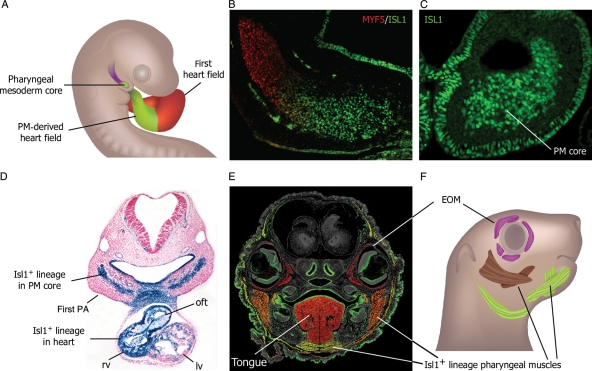
Isl1 marks pharyngeal mesoderm cells that populate the heart and the pharyngeal muscles. (A–C) Lineage studies in the chick have revealed the contribution of PM cells to the distal core of the pharyngeal arches and the heart. ISL1 protein expression is seen in this region (B–C), which is correlated with delayed differentiation of these myogenic progenitors (MYF5-). (D–F) Similarly, lineage analyses in mouse Isl1Cre; Rosa26LacZ E10.5 embryos (D) demonstrate the significant contribution of these cells to the core of the first pharyngeal arch and the heart. The heterogenic contribution of the Isl1 lineage to the head musculature at E16.5 is shown (E–F): A section of the Isl1Cre; Rosa26YFP embryo stained for muscle (red) or GFP (green). Isl1+ cells contribute strongly to lower jaw muscles (yellow), muscles of facial expression shown in the head periphery, and less to jaw closing muscles (orange). Extraocular muscles (EOMs) or tongue muscles are not derived from Isl1+ cells. (F) A diagram of the embryonic head (lateral view) reveals the contribution of high (yellow) and intermediate levels (brown) of the Isl1 lineage to the head musculature.
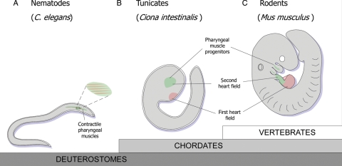
Isl1+ PM cells evolved from an ancestral myogenic programme for both cardiac and skeletal muscle lineages. (A) Nematodes such as the worm C. elegans do not have a heart; instead, they have a contractile pharyngeal muscle that functions like the heart in vertebrates. (B) Tunicates (Ciona intestinalis) are chordates that are considered as a ‘sister group’ to the vertebrates. Unlike nematodes, the heart and pharyngeal muscle cells in tunicates are seemingly distinct. Isl1+ cells in Ciona give rise to the pharyngeal muscles (termed siphon muscles), and not to the heart. (C) Reallocation of Isl1+ PM cells into the looping heart, which occurred during evolution from chordates to vertebrates, represents the emergence of the second heart field.
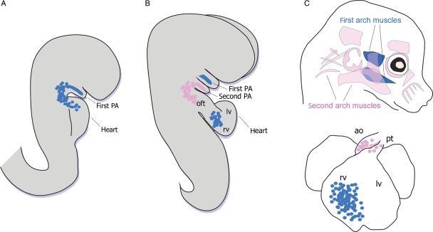
Clonal relationships of head and heart muscle progenitors in the mouse. Schematic representation of PM cells contributing to the core of the pharyngeal arches in the mouse embryo. (A) The linear heart tube at E8.0 is aligned at the level of the first pharyngeal arch; 1–2 days later, the heart undergoes looping, and shifts posteriorly. (B) First arch PM cells (blue) migrate to the developing heart tube to contribute to right ventricular myocardium. PM cells from the second arch (pink) migrate later (around E9.5–E10), to contribute to outflow tract myocardium (oft). (C) Diagram depicting the contribution of the different lineages to head muscles and heart myocardium: The first lineage (blue) contributes to masticatory muscles (temporalis and masseter) and to right ventricular (rv) myocardium. The second lineage (pink) contributes to facial expression muscles, and to myocardium at the base of the pulmonary trunk (pt) or aorta (ao). Modified after Lescroart F et al. Adapted with permission.
Similar articles
-
The contribution of Islet1-expressing splanchnic mesoderm cells to distinct branchiomeric muscles reveals significant heterogeneity in head muscle development.Development. 2008 Feb;135(4):647-57. doi: 10.1242/dev.007989. Epub 2008 Jan 9. Development. 2008. PMID: 18184728 Free PMC article.
-
Wnt signaling balances specification of the cardiac and pharyngeal muscle fields.Mech Dev. 2017 Feb;143:32-41. doi: 10.1016/j.mod.2017.01.003. Epub 2017 Jan 10. Mech Dev. 2017. PMID: 28087459 Free PMC article.
-
Collier/OLF/EBF-dependent transcriptional dynamics control pharyngeal muscle specification from primed cardiopharyngeal progenitors.Dev Cell. 2014 May 12;29(3):263-76. doi: 10.1016/j.devcel.2014.04.001. Epub 2014 May 1. Dev Cell. 2014. PMID: 24794633 Free PMC article.
-
Craniofacial Muscle Development.Curr Top Dev Biol. 2015;115:3-30. doi: 10.1016/bs.ctdb.2015.07.022. Epub 2015 Oct 1. Curr Top Dev Biol. 2015. PMID: 26589919 Review.
-
Emergence of heart and branchiomeric muscles in cardiopharyngeal mesoderm.Exp Cell Res. 2022 Jan 1;410(1):112931. doi: 10.1016/j.yexcr.2021.112931. Epub 2021 Nov 16. Exp Cell Res. 2022. PMID: 34798131 Review.
Cited by
-
Islet1-expressing cardiac progenitor cells: a comparison across species.Dev Genes Evol. 2013 Mar;223(1-2):117-29. doi: 10.1007/s00427-012-0400-1. Epub 2012 Apr 24. Dev Genes Evol. 2013. PMID: 22526874 Free PMC article. Review.
-
The impact of Drew Noden's work on our understanding of craniofacial musculoskeletal integration.Dev Dyn. 2022 Aug;251(8):1250-1266. doi: 10.1002/dvdy.471. Epub 2022 Apr 5. Dev Dyn. 2022. PMID: 35338756 Free PMC article. Review.
-
Heterogeneity of adult masseter muscle satellite cells with cardiomyocyte differentiation potential.Exp Cell Res. 2018 Oct 1;371(1):20-30. doi: 10.1016/j.yexcr.2018.05.028. Epub 2018 May 26. Exp Cell Res. 2018. PMID: 29842877 Free PMC article.
-
Nkx2.7 is a conserved regulator of craniofacial development.Nat Commun. 2025 Apr 23;16(1):3802. doi: 10.1038/s41467-025-58821-3. Nat Commun. 2025. PMID: 40268889 Free PMC article.
-
Integration of single-cell and spatial transcriptomics by SEU-TCA reveals the spatial origin of early cardiac progenitors.Genome Biol. 2025 Jun 10;26(1):158. doi: 10.1186/s13059-025-03633-3. Genome Biol. 2025. PMID: 40495257 Free PMC article.
References
-
- Psychoyos D, Stern CD. Fates and migratory routes of primitive streak cells in the chick embryo. Development. 1996;122:1523–1534. - PubMed
-
- Kinder SJ, Tsang TE, Quinlan GA, Hadjantonakis AK, Nagy A, Tam PP. The orderly allocation of mesodermal cells to the extraembryonic structures and the anteroposterior axis during gastrulation of the mouse embryo. Development. 1999;126:4691–4701. - PubMed
-
- Kelly RG, Brown NA, Buckingham ME. The arterial pole of the mouse heart forms from Fgf10-expressing cells in pharyngeal mesoderm. Dev Cell. 2001;1:435–440. - PubMed
-
- Mjaatvedt CH, Nakaoka T, Moreno-Rodriguez R, Norris RA, Kern MJ, Eisenberg CA, et al. The outflow tract of the heart is recruited from a novel heart-forming field. Dev Biol. 2001;238:97–109. - PubMed
