. 2011 Jun 3;286(22):19204-14.
doi: 10.1074/jbc.M111.224881. Epub 2011 Apr 15.

Structural and functional characterization of the Wnt inhibitor APC membrane recruitment 1 (Amer1)

Affiliations

Structural and functional characterization of the Wnt inhibitor APC membrane recruitment 1 (Amer1)

Kristina Tanneberger et al. J Biol Chem. .

Abstract

Amer1/WTX binds to the tumor suppressor adenomatous polyposis coli and acts as an inhibitor of Wnt signaling by inducing β-catenin degradation. We show here that Amer1 directly interacts with the armadillo repeats of β-catenin via a domain consisting of repeated arginine-glutamic acid-alanine (REA) motifs, and that Amer1 assembles the β-catenin destruction complex at the plasma membrane by recruiting β-catenin, adenomatous polyposis coli, and Axin/Conductin. Deletion or specific mutations of the membrane binding domain of Amer1 abolish its membrane localization and abrogate negative control of Wnt signaling, which can be restored by artificial targeting of Amer1 to the plasma membrane. In line, a natural splice variant of Amer1 lacking the plasma membrane localization domain is deficient for Wnt inhibition. Knockdown of Amer1 leads to the activation of Wnt target genes, preferentially in dense compared with sparse cell cultures, suggesting that Amer1 function is regulated by cell contacts. Amer1 stabilizes Axin and counteracts Wnt-induced degradation of Axin, which requires membrane localization of Amer1. The data suggest that Amer1 exerts its negative regulatory role in Wnt signaling by acting as a scaffold protein for the β-catenin destruction complex and promoting stabilization of Axin at the plasma membrane.


Figures

FIGURE 1.
FIGURE 1.
Schematic representation of Amer1 splice variants and deletion mutants analyzed in this paper. Amer1 is expressed as two splice variants, termed Amer1-S1 and Amer1-S2. The two N-terminal membrane localization domains (M1 and M2) are highlighted by light gray shading, the three APC interaction domains (A1–3) by dark gray shading, the central REA repeat domain involved in β-catenin binding by diagonal lines, and the regions involved in the interaction with Axin/Conductin by bold lines. Splicing of Amer1-S2 and the internal deletion of the REA repeat region in the Amer1ΔREA mutant are indicated by thin lines.
FIGURE 2.
FIGURE 2.
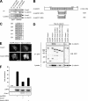
Amer1 interacts with β-catenin and recruits it to the plasma membrane. A, coimmunoprecipitation of endogenous Amer1 and β-catenin from lysates of HEK293T and SW480 cells. Immunoprecipitation (IP) was performed with mouse anti-β-catenin or control IgG antibodies. The asterisk indicates a nonspecific band. B, schematic representation of the β-catenin clones found in a yeast two-hybrid screen with Amer1(531–716) as a bait. The armadillo repeats are depicted as gray squares. Values represent β-galactosidase units of a representative yeast two-hybrid interaction analysis. C, amino acid sequence of the Amer1 REA repeat domain. REAH/Y motifs are highlighted in gray. D, Amer1 interacts with β-catenin via its REA repeat domain. Coimmunoprecipitation of Myc-tagged β-catenin with EGFP-tagged Amer1 or Amer1 deletion mutants in HEK293T cells (see Fig. 1). Note that full-size Amer1 and N-terminal fragments are expressed as two splice variants, Amer1-S1 and Amer1-S2, represented by the two bands on the Western blot analysis (see Fig. 1). E, Amer1 recruits β-catenin to the plasma membrane. Cotransfection of EGFP-tagged Amer1 or Amer1ΔREA (left panels, GFP fluorescence) with Myc-β-catenin (right panels, anti-β-catenin immunofluorescence) in MCF-7 cells. Scale bar = 20 μm. F, deletion of the REA repeat domain reduces the ability of Amer1 to repress a TCF/β-catenin-dependent transcriptional reporter. HEK293T cells stably expressing a β-catenin-responsive firefly luciferase were cotransfected with RFP-daLRP6 and the indicated EGFP-Amer1 constructs. Fold changes of luciferase activity are presented relative to control-transfected cells. Error bars indicate mean ± S.D. Similar expression of the daLRP6 and Amer1 constructs was verified by anti-LRP6 and anti-GFP Western blotting, respectively.
FIGURE 3.
FIGURE 3.
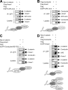
Amer1 acts as a scaffold for the β-catenin degradation complex. Protein interactions were determined by immunoprecipitation (IP) from transfected HEK293T cells. A, Amer1 can simultaneously interact with β-catenin and APC. Myc-β-catenin only coimmunoprecipitated with EGFP-APC-Arm when FLAG-Amer1 was present. B, Amer1 can simultaneously interact with APC and Conductin. FLAG-Conductin only coimmunoprecipitated with EGFP-APC-Arm when FLAG-Amer1 was present. C, Amer1 can simultaneously bind to Conductin and β-catenin. Myc-β-catenin only coimmunoprecipitated with EGFP-Conductin(455–782) when FLAG-Amer1 was present. D, APC and Conductin link β-catenin to Amer1. Coexpression of APC and FLAG-Conductin strongly increased the amount of Myc-β-catenin coimmunoprecipitated with EGFP-Amer1.
FIGURE 4.
FIGURE 4.
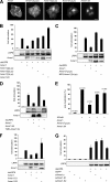
The membrane localization of Amer1 is required for repression of Wnt signaling. A, fluorescence micrographs of MCF-7 cells transfected with the EGFP-Amer1 constructs as indicated above the panels. Mutation of N-terminal lysine residues (Amer1(3μLys), Amer1(5μLys), and Amer1(7μLys)) or deletion of the membrane-targeting N terminus (Amer1ΔN, amino acids 207–1135) leads to the delocalization of Amer1 from the plasma membrane and its subsequent accumulation in the cytoplasm and nucleus. Note that Amer1 can appear as a diffuse staining or as discrete dots in the nucleus. Scale bar = 20 μm. B–F, HEK293T cells stably expressing a β-catenin-responsive firefly luciferase were cotransfected with RFP-daLRP6 and the indicated EGFP-Amer1 constructs. Fold changes of luciferase activity are presented relative to control-transfected cells. Error bars indicate mean ± S.D. Similar expression of the daLRP6 and Amer1 constructs was verified by anti-LRP6 and anti-GFP Western blotting, respectively. B, N-terminal lysine mutants of Amer1 are deficient in down-regulating the activity of a TCF/β-catenin-dependent transcriptional reporter. C, fusion of the Amer1(7μLys) mutant to a nuclear export sequence (NES-Amer1(7μLys)) does not restore inhibition of a TCF/β-catenin-dependent reporter. D, fusion of Amer1ΔN to the transmembrane domain of the low-density lipoprotein receptor (TMD-Amer1ΔN) restores inhibition of a TCF/β-catenin-dependent reporter. E, Amer1 mutants lacking membrane association are defective in down-regulating Wnt8-induced axis duplication in X. laevis embryos. The chart shows the normalized average percentage of axis induction from at least three independent experiments. Error bars indicate mean ± S.E. n, number of embryos injected. F and G, the membrane-bound splice variant Amer1-S1 represses Wnt signaling, whereas Amer1-S2 lacking the membrane targeting domain does not. Shown is the effect of overexpression (F) or knockdown (G) of Amer1 splice variants Amer1-S1 and Amer1-S2 on a TCF/β-catenin-dependent reporter upon activation with daLRP6.
FIGURE 5.
FIGURE 5.
Amer1 exerts its negative regulatory activity on Wnt signaling mainly under conditions of high cell density. Induction of Wnt target genes Conductin and LGR5 after knockdown of Amer1 as analyzed by RT-PCR or Western blotting (WB) analysis. siRNA-transfected HeLa cells were seeded at either high or low cell density and incubated with Wnt3A for 3.5 h (RT-PCR) or 5 h (WB) before cell lysis.
FIGURE 6.
FIGURE 6.
Amer1 increases the stability of Axin. A, knockdown of Amer1 reduces Axin levels. HEK293T or SW480 cells were transfected with the indicated siRNAs, and endogenous Axin was analyzed by Western blotting. The asterisk indicates a nonspecific band. B, knockdown of Amer1 using siRNA does not affect Axin mRNA levels as shown by RT-PCR analysis in HEK293T cells. C and D, overexpression of Amer1 increases Axin levels, which requires membrane localization (C) and Axin binding (D) of Amer1. The indicated EGFP-Amer1 constructs were coexpressed with FLAG-Axin in HEK293T cells. E and F, Amer1 increases the half-life of Axin. E, HEK293T cells were transfected with FLAG-Axin with or without EGFP-Amer1 and incubated with 100 μm cycloheximide (CHX) for the indicated time points. F, quantification of the experiment from E by densitometry. The levels of Axin protein at each time point were normalized to the starting level. G, overexpression of Amer1 prevents Wnt-induced down-regulation of Axin. HEK293T cells were transfected with the indicated EGFP-Amer1 constructs, incubated with Wnt3A for 4 h, and then endogenous Axin was analyzed by Western blotting. H, overexpression of Amer1 prevents down-regulation of Axin induced by daLRP6. HEK293T cells were transfected with FLAG-Axin, RFP-daLRP6, and EGFP-Amer1 as indicated. I, knockdown of Axin abolishes the negative regulatory effect of Amer1 on a TCF/β-catenin-dependent reporter. The TCF/β-catenin reporter assay was performed as described in Fig. 2F.

References

    1. Clevers H. (2006) Cell 127, 469–480 - PubMed
    1. Lustig B., Behrens J. (2003) J. Cancer Res. Clin. Oncol. 129, 199–221 - PubMed
    1. Angers S., Moon R. T. (2009) Nat. Rev. Mol. Cell Biol. 10, 468–477 - PubMed
    1. MacDonald B. T., Tamai K., He X. (2009) Dev. Cell 17, 9–26 - PMC - PubMed
    1. Huang H., He X. (2008) Curr. Opin. Cell Biol. 20, 119–125 - PMC - PubMed

Publication types

MeSH terms