

Isolevuglandins and mitochondrial enzymes in the retina: mass spectrometry detection of post-translational modification of sterol-metabolizing CYP27A1
- PMID: 21498512
- PMCID: PMC3121529
- DOI: 10.1074/jbc.M111.232546
Isolevuglandins and mitochondrial enzymes in the retina: mass spectrometry detection of post-translational modification of sterol-metabolizing CYP27A1
Abstract
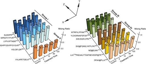
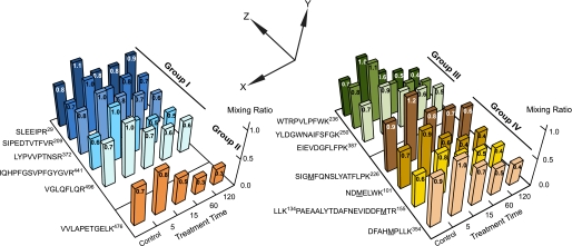
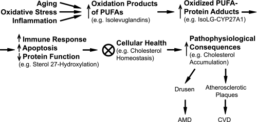
We report the first peptide mapping and sequencing of an in vivo isolevuglandin-modified protein. Mitochondrial cytochrome P450 27A1 (CYP27A1) is a ubiquitous multifunctional sterol C27-hydroxylase that eliminates cholesterol and likely 7-ketocholesterol from the retina and many other tissues. We investigated the post-translational modification of this protein with isolevuglandins, arachidonate oxidation products. Treatment of purified recombinant CYP27A1 with authentic iso[4]levuglandin E(2) (iso[4]LGE(2)) in vitro diminished enzyme activity in a time- and phospholipid-dependent manner. A multiple reaction monitoring protocol was then developed to identify the sites and extent of iso[4]LGE(2) adduction. CYP27A1 exhibited only three Lys residues, Lys(134), Lys(358), and Lys(476), that readily interact with iso[4]LGE(2) in vitro. Such selective modification enabled the generation of an internal standard, (15)N-labeled CYP27A1 modified with iso[4]LGE(2), for the subsequent analysis of a human retinal sample. Two multiple reaction monitoring transitions arising from the peptide AVLK(358)(-C(20)H(26)O(3))ETLR in the retinal sample were observed that co-eluted with the corresponding two (15)N transitions from the supplemented standard. These data demonstrate that modified CYP27A1 is present in the retina. We suggest that such protein modification impairs sterol elimination and likely has other pathological sequelae. We also propose that the post-translational modifications identified in CYP27A1 exemplify a general mechanism whereby oxidative stress and inflammation deleteriously affect protein function, contributing, for example, to cholesterol-rich lesions associated with age-related macular degeneration and cardiovascular disease. The proteomic protocols developed in this study are generally applicable to characterization of lipid-derived oxidative protein modifications occurring in vivo, including proteins bound to membranes.
Figures


Similar articles
-
Posttranslational modification by an isolevuglandin diminishes activity of the mitochondrial cytochrome P450 27A1.J Lipid Res. 2013 May;54(5):1421-9. doi: 10.1194/jlr.M035790. Epub 2013 Mar 11. J Lipid Res. 2013. PMID: 23479405 Free PMC article.
-
Abnormal vascularization in mouse retina with dysregulated retinal cholesterol homeostasis.J Clin Invest. 2012 Aug;122(8):3012-23. doi: 10.1172/JCI63816. Epub 2012 Jul 23. J Clin Invest. 2012. PMID: 22820291 Free PMC article.
-
Quantification of cholesterol-metabolizing P450s CYP27A1 and CYP46A1 in neural tissues reveals a lack of enzyme-product correlations in human retina but not human brain.J Proteome Res. 2011 Jan 7;10(1):241-8. doi: 10.1021/pr1008898. Epub 2010 Nov 16. J Proteome Res. 2011. PMID: 21049985 Free PMC article.
-
Isolevuglandin adducts in disease.Antioxid Redox Signal. 2015 Jun 20;22(18):1703-18. doi: 10.1089/ars.2014.6154. Epub 2015 Feb 18. Antioxid Redox Signal. 2015. PMID: 25557218 Free PMC article. Review.
-
Protein lipoylation: an evolutionarily conserved metabolic regulator of health and disease.Curr Opin Chem Biol. 2018 Feb;42:76-85. doi: 10.1016/j.cbpa.2017.11.003. Epub 2017 Nov 21. Curr Opin Chem Biol. 2018. PMID: 29169048 Free PMC article. Review.
Cited by
-
Pretreatment with pyridoxamine mitigates isolevuglandin-associated retinal effects in mice exposed to bright light.J Biol Chem. 2013 Oct 11;288(41):29267-80. doi: 10.1074/jbc.M113.498832. Epub 2013 Aug 22. J Biol Chem. 2013. PMID: 23970548 Free PMC article.
-
The Adductomics of Isolevuglandins: Oxidation of IsoLG Pyrrole Intermediates Generates Pyrrole⁻Pyrrole Crosslinks and Lactams.High Throughput. 2019 May 10;8(2):12. doi: 10.3390/ht8020012. High Throughput. 2019. PMID: 31083423 Free PMC article.
-
Mass spectrometry detection of isolevuglandin adduction to specific protein residues.Methods Mol Biol. 2015;1208:285-98. doi: 10.1007/978-1-4939-1441-8_21. Methods Mol Biol. 2015. PMID: 25323515 Free PMC article.
-
Lipid peroxidation generates biologically active phospholipids including oxidatively N-modified phospholipids.Chem Phys Lipids. 2014 Jul;181:1-33. doi: 10.1016/j.chemphyslip.2014.03.002. Epub 2014 Apr 2. Chem Phys Lipids. 2014. PMID: 24704586 Free PMC article. Review.
-
Brain Cytochrome P450: Navigating Neurological Health and Metabolic Regulation.J Xenobiot. 2025 Mar 14;15(2):44. doi: 10.3390/jox15020044. J Xenobiot. 2025. PMID: 40126262 Free PMC article. Review.
References
Publication types
MeSH terms
Substances
Grants and funding
LinkOut - more resources
Full Text Sources
