. 2011 Jun 10;286(23):20512-24.
doi: 10.1074/jbc.M111.237875. Epub 2011 Apr 15.

Competing interactions stabilize pro- and anti-aggregant conformations of human Tau

Affiliations

Competing interactions stabilize pro- and anti-aggregant conformations of human Tau

Susanne Wegmann et al. J Biol Chem. .

Abstract

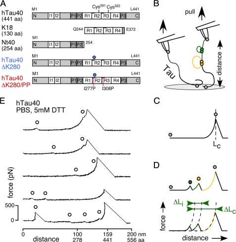
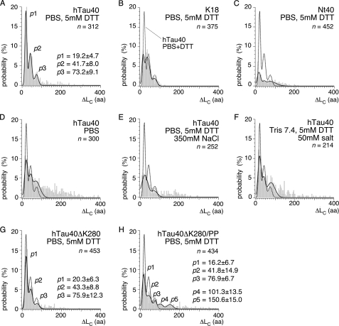
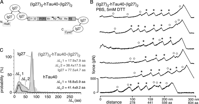
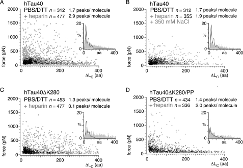
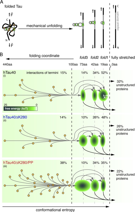
Aggregation of Tau into amyloid-like fibrils is a key process in neurodegenerative diseases such as Alzheimer. To understand how natively disordered Tau stabilizes conformations that favor pathological aggregation, we applied single-molecule force spectroscopy. Intramolecular interactions that fold polypeptide stretches of ~19 and ~42 amino acids in the functionally important repeat domain of full-length human Tau (hTau40) support aggregation. In contrast, the unstructured N terminus randomly folds long polypeptide stretches >100 amino acids that prevent aggregation. The pro-aggregant mutant hTau40ΔK280 observed in frontotemporal dementia favored the folding of short polypeptide stretches and suppressed the folding of long ones. This trend was reversed in the anti-aggregant mutant hTau40ΔK280/PP. The aggregation inducer heparin introduced strong interactions in hTau40 and hTau40ΔK280 that stabilized aggregation-prone conformations. We show that the conformation and aggregation of Tau are regulated through a complex balance of different intra- and intermolecular interactions.


Figures

FIGURE 1.
FIGURE 1.
Tau isoforms and SMFS setup. A, five investigated isoforms and constructs of the Tau protein. hTau40 (441 aa), the longest human isoform in the central nervous system containing four repeats (4R) and two N-terminal inserts (I1 and I2). K18 (130 aa), consisting of the hTau40 4R domain. Nt40, the 254-aa long N-terminal fragment of hTau40. hTau40ΔK280, a pro-aggregant mutant with a Lys280 deletion (blue circle) in the second repeat (R2) of hTau40. hTau40ΔK280/PP, an anti-aggregant mutant of hTau40ΔK280, in which Ile277 and Ile308 where exchanged against two prolines (red lines). B, for SMFS, Tau proteins adsorbed to amino-functionalized glass supports were picked up by the AFM tip and stretched by the AFM cantilever (probe) until their connection to the tip or the glass (gray closed circles in B-D) ruptured. Tau consists of unstructured protein regions (black lines) with intramolecular interactions (green and yellow filled circles) that fold peptide stretches of different lengths (yellow and green lines). Recording deflection and distance of the cantilever during consecutive approach-retract cycles provides force-distance (F-D) curves of single Tau molecules. C, stretching of a molecule having no intramolecular interactions results in a F-D curve that shows one major detachment peak at the contour length, LC, of the fully extended molecule. D, intramolecular interactions (green and yellow filled circles) can establish force barriers that are detected as additional force peaks in the F-D curve. For every additional force peak, the contour length relative to the detachment peak, ΔLC, and the rupture force was derived. The distance to the next rupture peak, ΔLi, in an F-D curve gives the length of the polypeptide stretch that unravels upon breaking an interaction. E, F-D curves recorded upon stretching single hTau40 molecules in PBS containing 5 mm DTT. The curves show a major detachment peak at the contour length of the Tau molecule (1 aa ≈ 0.36 nm) plus smaller force peaks at shorter contour lengths (open black circles) originating from intramolecular interactions.
FIGURE 2.
FIGURE 2.
ΔLC distribution of rupture peaks in hTau40, repeat domain construct K18, N-terminal fragment Nt40, and mutant proteins hTau40ΔK280 and hTau40ΔK280/PP. Contour lengths relative to the detachment peak, ΔLC, at which interactions were detected upon mechanically stretching hTau40 (A), K18 (B), and Nt40 (C) molecules in PBS containing 5 mm DTT. The most probable positions of rupture peaks were determined for each condition and construct from triple-Gaussian fits (solid lines in A–H) to the ΔLC distributions. For hTau40 in PBS/DTT (A), the most probable rupture peak positions were determined at p1 ∼19 aa, p2 ∼42 aa, and p3 ∼73 aa. D, contour lengths detected in hTau40 upon stretching in the absence of DTT (pure PBS); E, in buffer of 500 mm ionic strength (PBS/DTT + 350 mm NaCl); and F, in buffer of ∼50 mm ionic strength (Tris + 50 mm NaCl). G, contour lengths in the pro-aggregant mutant hTau40ΔK280; and H, the anti-aggregant mutant hTau40ΔK280/PP upon stretching in PBS + 5 mm DTT. The most probable positions of rupture peaks were determined from Gaussian fits (black lines) to the ΔLC distributions as p1 ∼20 aa, p2 ∼43 aa, and p3 ∼76 aa for hTau40ΔK280 (G), and as p1 ∼16 aa, p2 ∼42 aa, p3 ∼77 aa, p4 ∼101 aa, and p5 ∼151 aa for hTau40ΔK280/PP (H) (supplemental Table S1). n, gives the number of analyzed F-D curves.
FIGURE 3.
FIGURE 3.
SMFS of the (Ig27)3-hTau40-(Ig27)2 construct. A, fusion protein (Ig27)3-hTau40-(Ig27)2 in which hTau40 is flanked by three N-terminal and two C-terminal Ig27 domains. The full extension of hTau40 was guaranteed when detecting four or more Ig27 unfolding events in the F-D curve. B, F-D curves attained from stretching single (Ig27)3-hTau40-(Ig27)2 molecules in PBS containing 5 mm DTT show a mixture of Ig27 (*) and hTau40 (○) unfolding events. C, length distribution of unraveled polypeptide stretches, ΔLi, detected in F-D curves of (Ig27)3-hTau40-(Ig27)2 (gray) resemble that detected of isolated hTau40 (black). Triple Gaussian fit to the ΔLi distribution of (Ig27)3-hTau40-(Ig27)2 (gray line; n = 69) reveals interaction contour lengths of ΔLi1 ∼18 aa and ΔLi2 ∼38 aa of the sandwiched hTau40, plus the characteristic contour length of the Ig27 domains of ∼78 aa. A double Gaussian fit to the ΔLi distribution determined for hTau40 only (black line; n = 223) reveals most probable ΔLi of ΔLi1 ∼19 aa and ΔLi2 ∼41 aa. n, gives the number of analyzed F-D curves.
FIGURE 4.
FIGURE 4.
Heparin-induced interactions in hTau40, hTau40ΔK280, and hTau40ΔK280/PP. Scatter plots of rupture forces detected upon stretching hTau40 (A), hTau40ΔK280 (C), and hTau40ΔK280/PP (D) in PBS containing 5 mm DTT in the absence (black crosses in A and B, n = 312; C, n = 453; and D, n = 434; pulling velocity 875 nm/s) and presence (open gray circles in A, n = 477; C, n = 244; and D, n = 336; pulling velocity 1000 nm/s) of 0.33 mm heparin. Insets show the distributions of ΔLC in the presence (gray bars) of heparin. For comparison, the fits to the ΔLC distributions in the absence of heparin (black dashed lines) are shown. B, scatter plot of force peaks detected when stretching hTau40 in the presence of 0.33 mm heparin and ∼500 mm electrolyte concentrations (PBS + 5 mm DTT + 350 mm NaCl + 0.33 mm heparin; open gray circles (B, n = 355). Heparin binding induced a large number of high force interactions (300–1500 pN) over the full range of contour lengths in hTau40 (A) and hTau40ΔK280 (C). The ensemble of interactions in hTau40ΔK280/PP (D) showed only minor changes. Addition of heparin at ∼500 mm electrolyte reversed most of the heparin-induced interactions in B. n, gives the number of analyzed F-D curves.
FIGURE 5.
FIGURE 5.
Model of the unfolding energy landscape of full-length human Tau. A, scheme showing the main unfolding intermediates when stretching full-length Tau protein. Tau conformations (left) stabilized by interactions of the termini (orange circles) and the repeat domain (green circles) are forced into fully stretched conformations (right). Thereby, Tau unfolds stepwise taking the main unfolding intermediates stabilized by fold3, fold2, and fold1. B, schematic energy landscapes for the stretching of hTau40 (i), hTau40ΔK280 (ii), and hTau40ΔK280/PP (iii). The three main intramolecular interactions p1, p2, and p3 (green ellipses) establish energy wells in the unfolding landscape of Tau. The widths (x axis) of the three ellipses at 19 (p1), 42 (p2), and 73 aa (p3) estimate the widths xu of the energy wells stabilizing fold1, fold2, and fold3. The depth of every energy well, ΔG, is indicated by the color-coded scale bar. Different Tau conformations show different combinations of interactions in the termini and the repeat domain and stepwise unfold through various unfolding pathways (arrows in B, i, ii, and iii). Weak interactions of the termini break before the three abundant folds, fold1, fold2, and fold3, in the repeat domain. The larger number of unspecific, long peptide folds and the low stiffness of the fold3 interaction in hTau40ΔK280/PP (iii) may protect this Tau mutant from establishing aggregation conformations. In contrast, the ∼19- (fold1) and ∼42-aa (fold2) interactions are strengthened in hTau40ΔK280 (ii) and may thus be important for the aggregation of Tau.

Similar articles

Cited by

References

    1. Chien P., Weissman J. S., DePace A. H. (2004) Annu. Rev. Biochem. 73, 617–656 - PubMed
    1. Skrabana R., Sevcik J., Novak M. (2006) Cell Mol. Neurobiol. 26, 1085–1097 - PMC - PubMed
    1. Uversky V. N. (2002) Protein Sci. 11, 739–756 - PMC - PubMed
    1. Kidd M. (1963) Nature 197, 192–193 - PubMed
    1. Crowther R. A. (1990) Biochim. Biophys. Acta 1096, 1–9 - PubMed

Publication types

LinkOut - more resources