. 2011 Jun 10;286(23):20387-97.
doi: 10.1074/jbc.M111.236943. Epub 2011 Apr 15.

Visual insight into how low pH alone can induce actin-severing ability in gelsolin under calcium-free conditions

Affiliations

Visual insight into how low pH alone can induce actin-severing ability in gelsolin under calcium-free conditions

Renu Garg et al. J Biol Chem. .

Abstract

Gelsolin is a key actin cytoskeleton-modulating protein primarily regulated by calcium and phosphoinositides. In addition, low pH has also been suggested to activate gelsolin in the absence of Ca(2+) ions, although no structural insight on this pathway is available except for a reported decrement in its diffusion coefficient at low pH. We also observed ~1.6-fold decrease in the molecular mobility of recombinant gelsolin when buffer pH was lowered from 9 to 5. Analysis of the small angle x-ray scattering data collected over the same pH range indicated that the radius of gyration and maximum linear dimension of gelsolin molecules increased from 30.3 to 34.1 Å and from 100 to 125 Å, respectively. Models generated for each dataset indicated that similar to the Ca(2+)-induced process, low pH also promotes unwinding of this six-domain protein but only partially. It appeared that pH is able to induce extension of the G1 domain from the rest of the five domains, whereas the Ca(2+)-sensitive latch between G2 and G6 domains remains closed. Interestingly, increasing the free Ca(2+) level to merely ~40 nM, the partially open pH 5 shape "sprung open" to a shape seen earlier for this protein at pH 8 and 1 mm free Ca(2+). Also, pH alone could induce a shape where the g3-g4 linker of gelsolin was open when we truncated the C-tail latch from this protein. Our results provide insight into how under physiological conditions, a drop in pH can fully activate the F-actin-severing shape of gelsolin with micromolar levels of Ca(2+) available.


Figures

FIGURE 1.
FIGURE 1.
DLS data as a function of low pH and higher Ca2+ ions. Left, the relative change in diffusion coefficient of the gelsolin molecules shows how the molecular motion slows down by a factor of ∼1.6 upon sensing the lowering of pH from 9 to 5. The arrow indicates the pI of gelsolin (∼5.7). Right, this plot shows how the diffusion coefficient of gelsolin molecules decreases with increasing Ca2+ ion concentration in buffer.
FIGURE 2.
FIGURE 2.
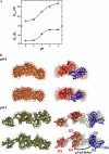
SAXS data analysis. A, low angle scattering intensity profiles from samples of gelsolin in buffers containing varying pH and their Guinier approximations (inset) are presented. B, Kratky plots of the SAXS data highlight the globular nature of the gelsolin molecules in solution. C, P(r) curves computed using the SAXS data show how the shape of the scattering species opens in buffers containing lower pH. D, the increase in the Dmax and RG values of the gelsolin as a function of pH as deduced from indirect Fourier transformation of the SAXS data are plotted.
FIGURE 3.
FIGURE 3.
Orthogonal views of the models restored for scattering molecules aid us in visualizing the opening up of the global structure of gelsolin as result of lower pH in buffers. Due to similarity in shape profiles, the low resolution models restored in pH 9, 8, and 7 were superimposed on the crystal structure of gelsolin under EGTA conditions (PDB ID: 1DON chain A). The models computed for gelsolin under pH 6 and 5 clearly provide structural evidence that the six-domain gelsolin molecules start unwinding at low pH.
FIGURE 4.
FIGURE 4.
Two rotated views of the models solved for gelsolin molecules under pH 8 (left) and 5 (right) are compared with structures known from x-ray crystallography to understand the solution structure of this protein under those conditions. The Cα traces of the N- (G1-G3) and C-(G4-G6) terminal halves of the molecule in the crystal structures are shown as red and blue tubes, respectively.
FIGURE 5.
FIGURE 5.
A, the increase in the Dmax values of the gelsolin molecules at pH 9–5 as a function of increasing Ca2+ ion concentration in buffer is presented. B, two rotated views of the chain-ensemble model solved for gelsolin molecules at pH 5 supplemented with 40 nm free Ca2+ are shown and compared with crystal structures of Ca2+-activated gelsolin halves to visualize the domain rearrangements in this protein upon sensing low pH and Ca2+ ions. The Cα traces of the N- and C-terminal halves of the molecule in the crystal structures are shown as red and blue tubes, respectively.
FIGURE 6.
FIGURE 6.
A, the increase in Dmax and RG values of the gelsolin protein truncated at the 693rd amino acid as a function of low pH alone is plotted here. B, two rotated views of the chain-ensemble models solved for C-terminal truncated gelsolin molecules under pH 8 (top) and 5 (bottom) under EGTA conditions are shown and compared with high resolution structures known from x-ray crystallography to affirm that pH alone can open the protein to its full extent if the G2/G6 interdomain contacts are removed. The Cα traces of the N- and C- terminal halves of the molecule in the crystal structures are shown as red and blue tubes, respectively.

Similar articles

Cited by

References

    1. Kwiatkowski D. J., Mehl R., Yin H. L. (1988) J. Cell Biol. 106, 375–384 - PMC - PubMed
    1. Ashish, Paine M. S., Perryman P. B., Yang L., Yin H. L., Krueger J. K. (2007) J. Biol. Chem. 282, 25884–25892 - PubMed
    1. Yin H. L., Stossel T. P. (1979) Nature 281, 583–586 - PubMed
    1. Yin H. L. (1987) Bioessays 7, 176–179 - PubMed
    1. Sun H. Q., Yamamoto M., Mejillano M., Yin H. L. (1999) J. Biol. Chem. 274, 33179–33182 - PubMed

Publication types