

Distinct roles of transforming growth factor-beta-activated kinase 1 (TAK1)-c-Rel and interferon regulatory factor 4 (IRF4) pathways in human T cell lymphotropic virus 1-transformed T helper 17 cells producing interleukin-9
- PMID: 21498517
- PMCID: PMC3122170
- DOI: 10.1074/jbc.M110.200907
Distinct roles of transforming growth factor-beta-activated kinase 1 (TAK1)-c-Rel and interferon regulatory factor 4 (IRF4) pathways in human T cell lymphotropic virus 1-transformed T helper 17 cells producing interleukin-9
Abstract
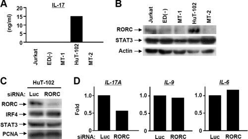
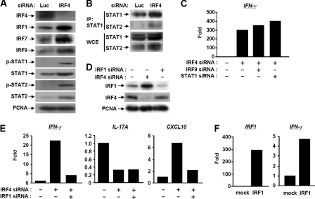
Investigation of helper T cell markers in HTLV-1-transformed cell lines demonstrated that HuT-102 has an IL-9-producing Th17 phenotype. We confirmed the vital role of retinoic acid-related orphan receptor C, a Th17 transcription factor, in the expression of IL-17. Interferon regulatory factor 4 (IRF4), a transcription factor overexpressed in all HTLV-1-infected cells, regulated IL-17 and IL-9 concomitantly. We further demonstrated a novel pathway for the regulation of Tax-induced cytokines, IL-9 and IL-6, through TAK1-mediated nuclear accumulation of c-Rel. A microarray analysis for IRF4 knocked down HuT-102 cells showed a significant up-regulation in the set of genes related to Th1, mainly IFN-γ and several transcription factors. T-bet and IRF1, but not STAT1 and IRF9, participated in counteracting the inhibitory effect of IRF4 on the production of IFN-γ. Finally, suppression of both IRF4 and c-Rel resulted in the reduced proliferation. Collectively, these findings indicate that TAK1-c-Rel and IRF4 pathways play distinct roles in the maintenance of IL-9-producing Th17 phenotype of HTLV-1-transformed cells.
Figures


Similar articles
-
Human T cell lymphotropic virus 1 manipulates interferon regulatory signals by controlling the TAK1-IRF3 and IRF4 pathways.J Biol Chem. 2010 Feb 12;285(7):4441-6. doi: 10.1074/jbc.M109.031476. Epub 2009 Dec 2. J Biol Chem. 2010. PMID: 19955181 Free PMC article.
-
Human T-cell leukemia virus type I Tax induces expression of the Rel-related family of kappa B enhancer-binding proteins: evidence for a pretranslational component of regulation.J Virol. 1991 Dec;65(12):6892-9. doi: 10.1128/JVI.65.12.6892-6899.1991. J Virol. 1991. PMID: 1719236 Free PMC article.
-
Interferon regulatory factor-4 activates IL-2 and IL-4 promoters in cooperation with c-Rel.Cytokine. 2011 Dec;56(3):564-72. doi: 10.1016/j.cyto.2011.08.014. Epub 2011 Sep 3. Cytokine. 2011. PMID: 21890374
-
Genes associated with T helper 17 cell differentiation and function.Front Biosci (Elite Ed). 2016 Jun 1;8(3):427-35. doi: 10.2741/E777. Front Biosci (Elite Ed). 2016. PMID: 27100349 Review.
-
Th9 and other IL-9-producing cells in allergic asthma.Semin Immunopathol. 2017 Jan;39(1):55-68. doi: 10.1007/s00281-016-0601-1. Epub 2016 Nov 17. Semin Immunopathol. 2017. PMID: 27858144 Review.
Cited by
-
Interleukin-6 mediates neutrophil mobilization from bone marrow in pulmonary hypertension.Cell Mol Immunol. 2021 Feb;18(2):374-384. doi: 10.1038/s41423-020-00608-1. Epub 2021 Jan 8. Cell Mol Immunol. 2021. PMID: 33420357 Free PMC article.
-
Interferon regulatory factor 4 as a therapeutic target in adult T-cell leukemia lymphoma.Retrovirology. 2020 Aug 28;17(1):27. doi: 10.1186/s12977-020-00535-z. Retrovirology. 2020. PMID: 32859220 Free PMC article.
-
Th17/IL-17 Axis in HTLV-1-Associated Myelopathy Tropical Spastic Paraparesis and Multiple Sclerosis: Novel Insights into the Immunity During HAMTSP.Mol Neurobiol. 2023 Jul;60(7):3839-3854. doi: 10.1007/s12035-023-03303-0. Epub 2023 Mar 22. Mol Neurobiol. 2023. PMID: 36947318 Review.
-
Downregulation of proinflammatory cytokines in HTLV-1-infected T cells by Resveratrol.J Exp Clin Cancer Res. 2016 Jul 22;35(1):118. doi: 10.1186/s13046-016-0398-8. J Exp Clin Cancer Res. 2016. PMID: 27448598 Free PMC article.
-
Prognostic impact of c-Rel nuclear expression and REL amplification and crosstalk between c-Rel and the p53 pathway in diffuse large B-cell lymphoma.Oncotarget. 2015 Sep 15;6(27):23157-80. doi: 10.18632/oncotarget.4319. Oncotarget. 2015. PMID: 26324762 Free PMC article.
References
-
- Osame M., Izumo S., Igata A., Matsumoto M., Matsumoto T., Sonoda S., Tara M., Shibata Y. (1986) Lancet. 2, 104–105 - PubMed
-
- Gessain A., Barin F., Vernant J. C., Gout O., Maurs L., Calender A., de Thé G. (1985) Lancet. 2, 407–410 - PubMed
-
- Hinuma Y., Komoda H., Chosa T. (1982) Int. J. Cancer. 29, 631–635 - PubMed
-
- Bazarbachi A., Ghez D., Lepelletier Y., Nasr R., de Thé H., El-Sabban M. E., Hermine O. (2004) Lancet. Oncol. 5, 664–672 - PubMed
-
- Grassmann R., Aboud M., Jeang K. T. (2005) Oncogene 24, 5976–5985 - PubMed
Publication types
MeSH terms
Substances
LinkOut - more resources
Full Text Sources
Molecular Biology Databases
Research Materials
Miscellaneous
