. 2011 Apr 15;25(8):789-94.
doi: 10.1101/gad.2027911.

Polycomb EZH2 controls self-renewal and safeguards the transcriptional identity of skeletal muscle stem cells

Affiliations

Polycomb EZH2 controls self-renewal and safeguards the transcriptional identity of skeletal muscle stem cells

Aster H Juan et al. Genes Dev. .

Abstract

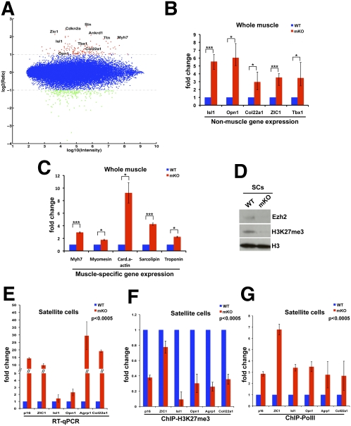
Satellite cells (SCs) sustain muscle growth and empower adult skeletal muscle with vigorous regenerative abilities. Here, we report that EZH2, the enzymatic subunit of the Polycomb-repressive complex 2 (PRC2), is expressed in both Pax7+/Myf5⁻ stem cells and Pax7+/Myf5+ committed myogenic precursors and is required for homeostasis of the adult SC pool. Mice with conditional ablation of Ezh2 in SCs have fewer muscle postnatal Pax7+ cells and reduced muscle mass and fail to appropriately regenerate. These defects are associated with impaired SC proliferation and derepression of genes expressed in nonmuscle cell lineages. Thus, EZH2 controls self-renewal and proliferation, and maintains an appropriate transcriptional program in SCs.


Figures

Figure 1.
Figure 1.
Ezh2 expression in limb muscles and SCs. (A) Limb muscle extracts from mice at P1–P60 probed with the indicated antibodies. (B) In P8 hindlimb muscles, EZH2 is expressed in Pax7+ cells. Arrowheads indicate cells positive for Pax7 and EZH2. Quantification of Pax7+/EZH2+ cells in P8 EDL muscles. (C, top panel) Myofiber-associated quiescent (0 h) Pax7+ SCs do not express a detectable level of EZH2. (Bottom panel) After 42 h, EZH2 is expressed in couplets of dividing Pax7+ SCs. (D) In single myofibers of Myf5-Cre/ROSA-YFP mice cultured for 42 h, EZH2 is expressed in both Pax7+/YFP (top panel) and Pax7+/YFP+ (bottom panel) cells. Arrows indicate YFP+ cells, and arrowheads indicate YFP cells. Bars, 50 μm.
Figure 2.
Figure 2.
Conditional Ezh2 ablation results in postnatal skeletal muscle defects and an impoverished SC pool. (A) RNA (left) and protein (right) analysis of Ezh2 in wild-type (WT) (Pax7-Cre−/−; Ezh2fl/fl) and mKO (Pax7-Cre+/−; Ezh2fl/fl) littermates. EZH2fl and EZH2Δ correspond to floxed and SET-deleted Ezh2 RNAs, respectively. (B,C) Both body and muscle mass are significantly reduced in P60 mKO compared with control (wild-type) littermates. Data are presented as mean ± SD (n = 6); (***) P < 0.0005; (**) P < 0.005. (D) H&E staining of transverse sections of P60 TA reveals a reduced myofiber CSA in Ezh2mKO animals (n = 3). Ninety-five percent confidence intervals do not overlap. (E) EDL single myofibers from adult (P60) Ezh2mKO animals have a reduced number of SCs. Arrows indicate Pax7+ cells. Data are presented as mean ± SD (n = 6); (*) P < 0.05. (F,G) Pax7 immunostaining of FACS-isolated wild-type and Ezh2mKO SCs. Arrows indicate cells stained positive for Pax7 and DAPI. Data are presented as mean ± SD (n = 3); (**) P < 0.005.
Figure 3.
Figure 3.
Skeletal muscle progenitors derived from Ezh2mKO animals have proliferative defects and precociously activate myogenin. (A,B) Immunostaining of H3K27me3 and Pax7 of P8 EDL. Arrowheads indicate cells stained positive for both Pax7 and H3K27me3. Arrows indicate Pax7+/H3K27me3 cells. Data are presented as mean ± SD (n = 3); (***) P < 0.0005; (**) P < 0.005. (C) Pax7 immunostaining of EDL single myofibers isolated from wild-type (WT) and Ezh2mKO mice cultured for 3 d. (*) P < 0.05. (D) FACS-isolated wild-type and Ezh2mKO SCs after 1 or 4 d in culture. Cells (1.5 × 10⁴) of wild-type and Ezh2 mKO SCs were seeded, and the total number obtained from counting cells present in 10 independent microscopic fields after 14, 48, and 96 h in culture was plotted in the curve graph (n = 4; P < 0.001 and P < 0.003 for 42-h and 96-h counts, respectively). (E) Percentage of FACS-isolated wild-type and Ezh2mKO SCs that have incorporated BrdU or that stained positive for histone-H3 (H3-P). Data are presented as mean ± SD for BrdU (n = 3; [**] P < 0.005) and H3-P (n = 4; [**] P < 0.005). (F) Increased p16 protein in Ezh2mKO muscles. GAPDH serves as a loading control. (G) Myogenin immunostaining of SC clones from wild-type and Ezh2mKO myofibers. Data are presented as mean ± SD; (*) P < 0.05.
Figure 4.
Figure 4.
Impaired muscle regeneration in Ezh2mKO animals. (A) Immunoblots of limb muscle extracts from wild-type (WT) or Ezh2mKO animals (n = 2) 3 d after CTX, probed with myogenin, Pax7, and GAPDH antibodies. (B) Pax7 and H3-P immunostaining of TA muscles from wild-type and Ezh2mKO animals 7 d after CTX injection. Data are presented as mean ± SD (n = 3); (***) P < 0.0005; (*) P < 0.05. (C) Embryonic MyHC [MyHC(emb)] immunostaining in wild-type and Ezh2mKO mice in 7-d regenerating myofibers. (D) H&E staining of transverse sections of wild-type or Ezh2mKO regenerating TA muscles 7 d after CTX injection indicates that the CSA of centrally nucleated myofibers is reduced in Ezh2mKO animals (n = 3). (Right panel) Ninety-five percent confidence intervals do not overlap.
Figure 5.
Figure 5.
Ezh2 maintains transcriptional identity of SCs. (A) Log intensity versus ratio scatter plot of gene expression obtained from microarray data derived from muscles of wild-type (n = 2) and Ezh2mKO (n = 2) animals. The X-axes represents the log10 of the average intensity of the data, and the Y-axes represents the log2 of their ratio. Each dot represents one gene. Up-regulated and down-regulated genes are color-coded red and green, respectively (fold change >2). (B,C) Expression of selected up-regulated genes identified in A by qPCR in muscles of wild-type or Ezh2mKO animals. Data are presented as mean ± SD (n = 3); (***) P < 0.0005; (*) P < 0.05. (D) EZH2 and H3K27me3 immunoblots of FACS-isolated Ezh2mKO SCs. Total histone H3 serves as loading control. (E) qPCR on selected up-regulated genes from FACS-isolated wild-type or Ezh2mKO SCs. Data are presented as mean ± SD (n = 3); P < 0.0005 for all the values. (F,G) ChIP-qPCR for H3K27me3 and PolII at selected genes up-regulated in Ezh2mKO SCs. Data are presented as mean ± SD (n = 3); P < 0.0005 for all the values.

Comment in

References

    1. Boyer LA, Plath K, Zeitlinger J, Brambrink T, Medeiros LA, Lee TI, Levine SS, Wernig M, Tajonar A, Ray MK, et al. 2006. Polycomb complexes repress developmental regulators in murine embryonic stem cells. Nature 441: 349–353 - PubMed
    1. Bracken AP, Kleine-Kohlbrecher D, Dietrich N, Pasini D, Gargiulo G, Beekman C, Theilgaard-Monch K, Minucci S, Porse BT, Marine JC, et al. 2007. The Polycomb group proteins bind throughout the INK4A–ARF locus and are disassociated in senescent cells. Genes Dev 21: 525–530 - PMC - PubMed
    1. Buckingham M, Relaix F 2007. The role of Pax genes in the development of tissues and organs: Pax3 and Pax7 regulate muscle progenitor cell functions. Annu Rev Cell Dev Biol 23: 645–673 - PubMed
    1. Caretti G, Di Padova M, Micales B, Lyons GE, Sartorelli V 2004. The Polycomb Ezh2 methyltransferase regulates muscle gene expression and skeletal muscle differentiation. Genes Dev 18: 2627–2638 - PMC - PubMed
    1. Chamberlain SJ, Yee D, Magnuson T 2008. Polycomb repressive complex 2 is dispensable for maintenance of embryonic stem cell pluripotency. Stem Cells 26: 1496–1505 - PMC - PubMed

Publication types

MeSH terms

Substances