. 2011 Apr 15;25(8):863-74.
doi: 10.1101/gad.2016411.

The initial phase of chromosome condensation requires Cdk1-mediated phosphorylation of the CAP-D3 subunit of condensin II

Affiliations

The initial phase of chromosome condensation requires Cdk1-mediated phosphorylation of the CAP-D3 subunit of condensin II

Satoshi Abe et al. Genes Dev. .

Abstract

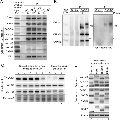
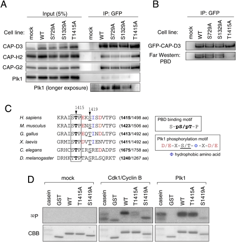
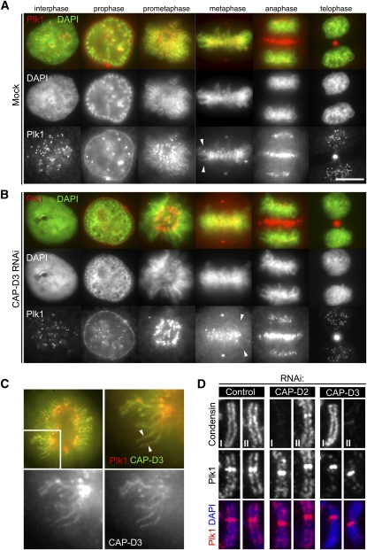
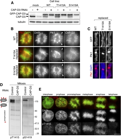
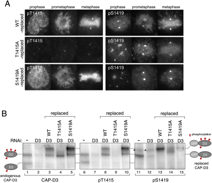
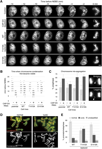
The cell cycle transition from interphase into mitosis is best characterized by the appearance of condensed chromosomes that become microscopically visible as thread-like structures in nuclei. Biochemically, launching the mitotic program requires the activation of the mitotic cyclin-dependent kinase Cdk1 (cyclin-dependent kinase 1), but whether and how Cdk1 triggers chromosome assembly at mitotic entry are not well understood. Here we report that mitotic chromosome assembly in prophase depends on Cdk1-mediated phosphorylation of the condensin II complex. We identified Thr 1415 of the CAP-D3 subunit as a Cdk1 phosphorylation site, which proved crucial as it was required for the Polo kinase Plk1 (Polo-like kinase 1) to localize to chromosome axes through binding to CAP-D3 and thereby hyperphosphorylate the condensin II complex. Live-cell imaging analysis of cells carrying nonphosphorylatable CAP-D3 mutants in place of endogenous protein suggested that phosphorylation of Thr 1415 is required for timely chromosome condensation during prophase, and that the Plk1-mediated phosphorylation of condensin II facilitates its ability to assemble chromosomes properly. These observations provide an explanation for how Cdk1 induces chromosome assembly in cells entering mitosis, and underscore the significance of the cooperative action of Plk1 with Cdk1.


Figures

Figure 1.
Figure 1.
Regulation of condensin II in mitosis. (A) Immunoprecipitation of Plk1 with condensin II. Condensin II components were immunoprecipitated from a chromosome-enriched fraction prepared from mitotic cell extracts using either of two different antibodies to CAP-D3 (#1 or #2), CAP-G2, CAP-H2, or nonimmune IgG as a control, and resulting precipitates (IP) were analyzed by immunoblotting with the antibodies indicated. Note that none of the condensin I subunits copurified with the condensin II complex, discounting the possibility that the interaction between condensin II and Plk1 is mediated indirectly by DNA. (B) Plk1 binds directly to CAP-D3 in a phosphorylation-dependent manner. Immunoprecipitates with either control or CAP-D3 antibodies from mitotic cell extracts were incubated with or without phosphatase (PPase), and were analyzed by immunoblotting with indicated antibodies (left panel) and Far-Western blotting with PBD (right panel). (C) Detection of CAP-D3 phosphorylation in mitotic cell extracts. Synchronized cell populations were analyzed by immunoblotting at the indicated times after the release from thymidine arrest (i.e., early S phase) (lanes 1–6) or nocodazole arrest (i.e., mitosis) (lanes 7,8). As reported previously (Yeong et al. 2003; Lipp et al. 2007), slowly migrating bands of CAP-D3 were detected specifically during mitosis, but little retardation was detectable for CAP-G2 and CAP-H2. (D) Mitotic phosphorylation of condensin II depends on Cdk1 and Plk1. Thymidine-arrested interphase cells (lane 1), nocodazole-arrested mitotic cells (lane 2), a Cdk1 inhibitor RO3306-treated nocodazole-arrested mitotic cell (lane 3), or a Plk1 inhibitor BI2536-treated nocodazole-arrested mitotic cell (lane 4) were analyzed by immunoblotting for condensin subunits. The mobility shifts for Cdc27 verify that these pretreatments efficiently abolished the activities of the targeted kinases in mitotic cells (Kraft et al. 2003).
Figure 2.
Figure 2.
Identification of phosphorylation sites on CAP-D3. (A) Immunoprecipitation of Plk1 with CAP-D3 is abolished in a Thr 1415 nonphosphorylatable mutant. Mitotic cell extracts prepared from cells stably expressing the indicated version of GFP-tagged CAP-D3 were subjected to immunoprecipitation analysis with antibodies to GFP and were immunoblotted with the antibodies indicated. (B) The binding of PBD to CAP-D3 is lost in the T1415A mutant. The indicated series of GFP-tagged CAP-D3 proteins were immunoprecipitated (top panel) and analyzed by Far-Western analysis with PBD (bottom panel). Note that, like the endogenous protein, GFP-CAP-D3 can be detected as two major bands in mitotic extracts from the wild-type and S729A and S1329A mutant cells, but not the T1415A cells. (C) A PBD-binding motif in CAP-D3. Among three candidate sites, Thr 1415 and Ser 1419 fall within a PBD-binding motif (boxed) and the Plk1 consensus phosphorylation site (color-coded), respectively. Equivalent regions from orthologous proteins from different species are aligned, highlighting the evolutionary conservation of these motifs. The numbers in brackets indicate positions of the Thr 1415-equivalent threonine (bold) in the full amino acid length of CAP-D3 protein. (D) Cdk1 and Plk1 mediate phosphorylation of Thr 1415 and Ser 1419, respectively, in vitro. A series of polypeptides corresponding to a partial fragment of CAP-D3 that encompasses the prospective phosphorylation sites were incubated with mock (control), Cdk1/Cyclin B, or Plk1. (Top panels) Incorporation of 32P was detected by autoradiography. (Bottom panels) Coomassie Brilliant Blue staining (CBB) verifies that equivalent amounts of substrate appear in each lane.
Figure 3.
Figure 3.
Enrichment of Plk1 on chromosomal axes. (A) Localization of Plk1 by immunofluorescence microscopy. Exponentially growing HeLa cells were fixed with methanol, incubated with antibodies to Plk1, and labeled with an Alexa fluorescent dye (red). DNA was stained with DAPI (green). Representative cells in interphase, prophase, prometaphase, metaphase, anaphase, and telophase are shown. Note the chromosome axial staining with Plk1 antibodies in prometaphase and metaphase (arrowheads). Bar, 10 μm. (B) Enrichment of Plk1 on chromosome axes is lost in the absence of CAP-D3. Cells depleted of CAP-D3 were processed for immunofluorescence microscopy as in A. Note that Plk1 signals on chromosome axes are displaced (arrowheads), while the other localizations are preserved. (C) Colocalization of Plk1 with CAP-D3. Wild-type GFP-CAP-D3-expressing cells were fixed and stained with Plk1 antibodies. Both CAP-D3 (right bottom panel; green in top merged panels) and Plk1 (left bottom panel; red in top merged panels) signals are visible on chromosome axes. (D) Enrichment of Plk1 at chromosome axes depends on condensin II but not condensin I. Chromosome spread samples prepared from mitotic cells that had been depleted of CAP-D2, CAP-D3, or mock (control) were fixed and stained for Plk1 with either condensin I (CAP-H) or condensin II (CAP-D3), as indicated. Quantification of fluorescence intensities for Plk1 signals is shown in Supplemental Figure 5A.
Figure 4.
Figure 4.
Mitotic phosphorylation of CAP-D3 on Thr 1415 and Ser 1419. (A) Replacement of endogenous CAP-D3 protein with GFP-tagged CAP-D3 proteins. HeLa cells that stably express GFP-tagged GFP-D3, either wild-type (WT) or the nonphosphorylatable mutant for Thr 1415 (T1415A) or Ser 1419 (S1419A), were generated. Expression of endogenous CAP-D3 was suppressed by RNAi designed to target the untranslated region of the gene. Interphase cell extracts were analyzed by immunoblotting for the amount of endogenous CAP-D3 (bottom bands) and GFP-CAP-D3 (top bands). (B) Delocalization of Plk1 from chromosome axes in T1415A-replaced cells. Wild-type-replaced, T1415A-replaced, or S1419A-replaced cells were fixed and stained with Plk1 antibodies (red). DNA was stained with DAPI (green). Representative prometaphase cells are shown. Note that T1415A and S1419A were both found enriched at chromosomal axes at levels comparable with the wild-type version, which discounts the possibility that these phosphorylations have a major role in the chromosomal association of condensin II. (C) Enrichment of Plk1 at chromosome axes is perturbed in T1415A-replaced cells. Fixed chromosome spread samples prepared from mitotic cells that had been replaced with the indicated version of CAP-D3 were stained with Plk1 antibodies. Quantification of fluorescence intensities for Plk1 signals is shown in Supplemental Figure 5B. (D) Generation of phospho-specific CAP-D3 antibodies. Total extracts of nocodazole-arrested cells treated with either mock (control) or CAP-D3 RNAi were subjected to immunoblotting by pT1415 or pS1419 antibodies. Note that two major pT1415 reactive bands and one major pS1419 reactive band (as denoted on the left side) are diminished in CAP-D3-depleted cells. Bands that do not disappear after CAP-D3 depletion are nonspecific reactive proteins (a band marked by an asterisk, for example). Specific reactivity to phospho-sites and mitotic forms are later shown in Supplemental Figure 6, A and B, respectively. (E) Timing of CAP-D3 Thr 1415 phosphorylation during mitotic progression. Fixed HeLa cells were stained with pT1415 antibodies and labeled with an Alexa dye (red). DNA was stained with DAPI (green). Representative pictures of interphase, prophase, prometaphase, metaphase, and early and late anaphases are shown. Bar, 10 μm. For p1419 staining, see Supplemental Figure 6B.
Figure 5.
Figure 5.
Hyperphosphorylation of CAP-D3 depends on CAP-D3-bound Plk1. (A) Reduced levels of Ser 1419 phosphorylation in T1415A-replaced cells. Cells in which endogenous CAP-D3 was replaced by either wild-type (top panels), T1415A (middle panels), or S1419A (bottom panels) forms of GFP-CAP-D3 were fixed and stained with pT1415 or pS1419 antibodies. Note that pS1419 antibodies cross-react with an unidentified epitope at centrosomes, seen as two marked dots that do not disappear after depletion of CAP-D3. Quantification of fluorescence intensities is summarized in Supplemental Figure 5, C and D. (B) Regulation of Thr 1415 and Ser 1419 phosphorylation. (First two lanes of each panel) Two species with different phosphorylation levels of endogenous CAP-D3 can be detected in mitotic cell extracts that diminish after CAP-D3 depletion, as depicted on the left side. (Last three lanes of each panel) Mitotic extracts from wild-type-replaced, T1415A-replaced or S1419A-replaced cells were analyzed. As replaced CAP-D3 proteins are tagged with GFP, they migrate slower than the endogenous proteins. The two corresponding phosphorylated forms of GFP-CAP-D3 are positioned on the right side. Asterisks mark nonspecific bands, which do not appear in immunopurified GFP-CAP-D3 samples (Supplemental Fig. 6A).
Figure 6.
Figure 6.
Cdk1-mediated phosphorylation of Thr 1415 is required for the full phosphorylation of condensin II. (A) Mitotic phosphorylation of non-Smc subunits of condensin II is perturbed in the T1415A mutant. Mitotic cell extracts prepared from indicated cell lines, with or without RNAi to Plk1 or to CAP-D3, were analyzed with the antibodies to condensin subunits. Parental cell line indicates the cell population that does not express any tagged protein. Note that changes in phosphorylation levels, as seen for condensin II subunits, are not readily detectable for condensin I subunits. (B) Phosphorylation of Thr 1415 and Ser 1419 depends primarily on Cdk1 and Plk1, respectively. Total cell extracts were prepared from thymidine-arrested interphase cells (lane 1) or nocodazole-arrested mitotic cells (lanes 2–4) in which the activity of Cdk1 or Plk1 is inhibited by RO3306 or BI2536 treatment, respectively (lanes 3,4), and were analyzed by immunoblotting using the antibodies indicated. Note that pT1415 and pS1419 antibodies can hardly detect CAP-D3 protein in interphase cells (cf. lanes 1 and 2), indicating the specific reactivity of these antibodies to mitotic phosphorylated forms. (C) Illustrations depicting how condensin II complex is phosphorylated in mitosis, and how phosphorylations are affected in the T1415A and S1419A mutants. The model predicts the crucial role of Cdk1 in phosphorylating CAP-D3 at Thr 1415, which triggers the full phosphorylation of the condensin II complex, and explains why the phosphorylation levels are markedly decreased in T1415A-replaced but not S1419A-replaced cells.
Figure 7.
Figure 7.
Nonphosphorylatable CAP-D3 mutants defective in mitotic functions of condensin II. (A) Analysis of the initial phases of chromosome condensation in live cells. Prophase image sequences were aligned on the time axis according to time before NEBD, which is defined by loss of a defined nuclear boundary. Bar, 5 μm. (B) Quantification of chromosome condensation in prophase. Time (minutes before NEBD) when chromosome condensation first became recognizable was scored and plotted for the indicated live-cell recordings. (C) Analysis of defective chromosome segregation in anaphase in nonphosphorylatable CAP-D3 mutants. During live-cell imaging analysis, anaphase cells were assessed for the presence of lagging and/or bridging chromosomes, as exemplified in the panels on the right. Mean ± SD from three experiment replicates (n = 20∼30 cells per experiment) are shown in the histogram. Similar results were obtained in a fixed-cell analysis (Supplemental Fig. 8). (D) Examples of chromosome spreads demonstrating normal (left panels) or curly (right panels) appearance. Chromosome spreads were prepared from cells that had been treated with a hypotonic buffer and stained with CAP-H (a condensin I subunit) antibodies (red). DNA was counterstained with DAPI (green). (E) Abnormal curly change of chromosomal axes in nonphosphorylatable CAP-D3 mutants. More than 100 cells per indicated condition were examined and scored based on their chromosomal axis appearance (mean ± SD).

References

    1. Archambault V, Glover DM 2009. Polo-like kinases: conservation and divergence in their functions and regulation. Nat Rev Mol Cell Biol 10: 265–275 - PubMed
    1. Elia AE, Rellos P, Haire LF, Chao JW, Ivins FJ, Hoepker K, Mohammad D, Cantley LC, Smerdon SJ, Yaffe MB 2003. The molecular basis for phosphodependent substrate targeting and regulation of Plks by the Polo-box domain. Cell 115: 83–95 - PubMed
    1. Gerlich D, Hirota T, Koch B, Peters JM, Ellenberg J 2006. Condensin I stabilizes chromosomes mechanically through a dynamic interaction in live cells. Curr Biol 16: 333–344 - PubMed
    1. Giet R, Glover DM 2001. Drosophila aurora B kinase is required for histone H3 phosphorylation and condensin recruitment during chromosome condensation and to organize the central spindle during cytokinesis. J Cell Biol 152: 669–682 - PMC - PubMed
    1. Gimenez-Abian JF, Clarke DJ, Mullinger AM, Downes CS, Johnson RT 1995. A postprophase topoisomerase II-dependent chromatid core separation step in the formation of metaphase chromosomes. J Cell Biol 131: 7–17 - PMC - PubMed

Publication types

MeSH terms

LinkOut - more resources