. 2011 Apr 15;25(8):875-88.
doi: 10.1101/gad.622411.

Phosphorylation-dependent regulation of the F-BAR protein Hof1 during cytokinesis

Affiliations

Phosphorylation-dependent regulation of the F-BAR protein Hof1 during cytokinesis

Franz Meitinger et al. Genes Dev. .

Abstract

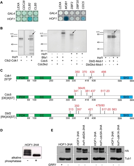
Spatial and timely coordination of cytokinesis is crucial for the maintenance of organelle inheritance and genome integrity. The mitotic exit network (MEN) pathway controls both the timely initiation of mitotic exit and cytokinesis in budding yeast. Here we identified the conserved F-BAR protein Hof1 as a substrate of the MEN kinase complex Dbf2-Mob1 during cytokinesis. We show that polo-like kinase Cdc5 first phosphorylates Hof1 to allow subsequent phosphorylation by Dbf2-Mob1. This releases Hof1 from the septin ring and facilitates Hof1 binding to the medial actomyosin ring (AMR), where Hof1 promotes AMR contraction and membrane ingression. Domain structure analysis established that the central, unstructured, region of Hof1, named the ring localization sequence (RLS), is sufficient to mediate Hof1's binding to the medial ring in a cell cycle-dependent manner. Genetic and functional data support a model in which Dbf2-Mob1 regulates Hof1 by inducing domain rearrangements, leading to the exposure of the Hof1 RLS domain during telophase.


Figures

Figure 1.
Figure 1.
Domain analysis of Hof1 bud neck localization. (A) Schematic representation (numbers represent amino acid positions) and quantification of metaphase- and anaphase-arrested cells (percentage) carrying the indicated Hof1 constructs. (FCH) FER/CIP4 homology; (CC) coiled coil; (SH3) SRC homology 3; (P) PEST sequence. “0” means no localization at the indicated bud neck region, yet protein levels were not decreased in comparison with wild-type Hof1 (see Supplemental Fig. S1). The cartoons represent the bud neck region showing “strong” and “weak” Hof1-GFP signals associated with septins (double bars) or at the medial ring (single bar). (B,D) Representative examples of Hof1-GFP signals at the bud neck in metaphase-arrested (B) and anaphase-arrested (D) cells, taken from the indicated cell backgrounds. (C) Quantification of Hof1-GFP fluorescence intensity at the bud neck (in arbitrary units) in metaphase-arrested cells (n = 15 per strain). (E) Schematic representation of the RLS region (pink) and a representative cell expressing HOF1-300–500-GFP.
Figure 2.
Figure 2.
The F-BAR domain and RLS are important for Hof1 function at the bud neck. (A) Electron micrographs of HOF1 and hof1Δ cells. The number of inspected cells is indicated in brackets. (B–F) Time-lapse series of INN1-GFP expressed in the indicated cell backgrounds. The first panel of each series shows a phase-contrast image. Cell boundaries are outlined. The bottom panels show the enlargement of the bud neck region outlined by the square. Arrowheads in enlarged images mark the boundary of the bud neck. (G) Quantification of B–F. (H) Quantification of the duration of Inn1-GFP at the bud neck of B–F. t-test: (*) t < 0.01; (**) t < 0.001.
Figure 3.
Figure 3.
Hof1 is a substrate of the Clb2–Cdk1, Cdc5, and Dbf2–Mob1 kinases. (A) Yeast two-hybrid interaction between LexA and Gal4 gene fusions, as indicated. Development of a blue color indicates interaction. (B) Autoradiographs showing Hof1 phosphorylated by purified Clb2–Cdk1, Cdc5, and Dbf2–Mob1 in vitro (arrows), but not by the corresponding kinase-dead (kd) mutants. Asterisks mark the autophosphorylation of Clb2–Cdk1 and Dbf2–Mob1. Bfa1 served as positive control for Cdc5 activity. (C) Schematic representation of Hof1 phosphorylation sites identified by MS for Clb2–Cdk1, Cdc5, and Dbf2–Mob1 in vitro (depicted in red). Consensus sites are indicated for each kinase. Numbers denote amino acids positions. Hof1 domains are represented as in Figure 1A. (D) Immunoblot using anti-HA antibodies showing immunoprecipitated Hof1-3HA treated with alkaline phosphatase (+) or buffer (−). (E) Comparison analysis of the phosphorylation pattern of indicated Hof1-3HA constructs expressed in Gal1-GRR1 cells upon depletion of GRR1 by glucose addition (GRR1). Protein levels of Hof1-3HA in cells expressing GRR1 (GRR1+) are shown for comparison. Hof1-3HA was detected using anti-HA antibodies. Tubulin (Tub1) served as loading control.
Figure 4.
Figure 4.
Hof1 phosphorylation is important for its function. (A) Schematic representation of Hof1 (as in Fig. 1A) showing the position of essential phosphorylation sites (depicted in red). (B) Serial dilutions of cyk3Δ hof1Δ URA3-CYK3 cells carrying the indicated LEU2-based plasmids. Cells were spotted on SC-complete and 5-FOA plates. Only URA3-negative cells grow on 5-FOA; i.e., no growth denotes growth lethality upon loss of URA3-based CYK3 plasmid. (C) Yeast two-hybrid interactions between the indicated LEXA and GAL4 gene fusions. Development of a blue color indicates interaction. (D) Quantification of C; β-galactosidase units are given in arbitrary units. (E) Pull-down assay using purified Dbf2–Mob1-GST complexes (immobilized on sepharose beads) incubated with yeast cell lysates of the indicated cell backgrounds. Immunoblots were detected using anti-HA (low and high exposure times are shown) and anti-GST antibodies. Signal intensities (arbitrary units) and the ratio of Hof1-3HA or Hof1-S517A-3HA bound to Mob1-GST are indicated.
Figure 5.
Figure 5.
Hof1 colocalizes with Cdc5 and Dbf2–Mob1 at the bud neck. Cycling cells of GFP-DBF2 HOF1-mCherry (A), GFP-DBF2 Cyk3-mCherry (B), CDC5-GFP HOF1-mCherry (C), and CDC5-GFP CYK3-mCherry (D) were analyzed by fluorescence microscopy. The merged images in A–D represent enlargements of the bud neck area. Please note that GFP-Dbf2 also localizes at the SPBs (dot-like signals). (E) Overview of the timely localization of Hof1, Cdc5, Dbf2–Mob1, and Cyk3 at the bud neck. Steps 1–6 are exemplified by cells shown in A–D. Hof1 localizes at the bud neck (septin-like staining) prior to Dbf2–Mob1 (A, panel 1), and relocalizes to a single medial ring concomitantly with the recruitment of Dbf2–Mob1 to the bud neck (A, panels 3–5). (A, panel 6) Hof1 contracts with the AMR, whereas Dbf2 does not. (B, panels 4,5) The late cytokinetic marker Cyk3 gets recruited to the bud neck after Dbf2. (B, panel 6) Cyk3 contracts with the AMR, whereas Dbf2 stays as a ring at the bud neck. Hof1 colocalizes with Cdc5 at the septin scaffold (C, panel 2), but Cdc5 is not part of the contractile AMR, since it never colocalizes with Cyk3 (D, panels 2,5).
Figure 6.
Figure 6.
Phosphorylation of Hof1 induces its rearrangement at the bud neck. (A) Localization of Hof1-GFP and Hof1–dbf2E-GFP in dbf2-2 dbf20Δ cells arrested for 3 h at 37°C. (B) Immunoblots show the levels of Hof1-GFP and Hof1–dbf2E-GFP, as indicated. Tub1 served as loading control. (C) Two-hybrid interaction between the indicated LexA and Gal4 fusion proteins. (D) Quantification of C representing β-galactosidase assays of three independent experiments. The graph shows the relative interaction of Hof1–dbf2A and Hof1–dbf2E with Cdc10 and Mob1. Hof1–Mob1 and Hof1–Cdc10 interactions were normalized to 1. (E) Specific bud neck localization of Hof1-GFP constructs in metaphase-arrested cells was performed as in Figure 1. (F) Quantification of E. Fluorescence intensity is given in arbitrary units (please note that the experiments shown in E and F were done together with the experiments shown in Fig. 1; i.e., the values for Hof1 and Hof1-W637A are the same in both figures). (G) Specific bud neck localization of Hof1-GFP constructs in anaphase-arrested cells was performed as in Figure 1. (H) Localization of the indicated Hof1-GFP constructs in dbf2-2 dbf20Δ cells arrested for 3 h at 37°C. Enlargements of the bud neck region are shown in A and H.
Figure 7.
Figure 7.
Hof1 phosphomimetic mutations in Dbf2 sites partially rescue dbf2-2 dbf20Δ-dependent cytokinesis defects. Cells of the indicated genotypes were arrested in G1 at 23°C with α-factor. Cells were released in nocodazole at 37°C to promote metaphase arrest and inactivation of Dbf2-2. After release from metaphase, overexpression of SIC1 was induced by addition of galactose to the culture medium to overcome the mitotic exit defects of dbf2-2 dbf20Δ cells. Samples were taken every 30 min to monitor Hof1 localization and actin repolarization. One representative cell (taken from time point 60 min) is shown in A–D. The levels of Hof1-GFP, Sic1, and Clb2 were monitored by immunoblotting using anti-GFP, anti-Sic1, and anti-Clb2 antibodies. Tub1 served as a loading control. The graphs indicate the percentage of cells (n = 100–150) showing actin repolarization at the bud neck and Hof1-GFP at septins, at the AMR ring, or as a contracted ring.
Figure 8.
Figure 8.
Model for Hof1 regulation by phosphorylation. The F-BAR and SH3 domains contribute to Hof1's association with septins during most phases of the cell cycle. In late mitosis, the specific association of Hof1 with the septin scaffold gets resolved after phosphorylation of Hof1 by Dbf2–Mob1. Phosphorylated Hof1 binds to the medial ring and, together with Cyk3 and Inn1, coordinates AMR contraction with primary septum formation.

Similar articles

Cited by

References

    1. Asano S, Park JE, Sakchaisri K, Yu LR, Song S, Supavilai P, Veenstra TD, Lee KS 2005. Concerted mechanism of Swe1/Wee1 regulation by multiple kinases in budding yeast. EMBO J 24: 2194–2204 - PMC - PubMed
    1. Aspenstrom P 2009. Roles of F-BAR/PCH proteins in the regulation of membrane dynamics and actin reorganization. Int Rev Cell Mol Biol 272: 1–31 - PubMed
    1. Aspenstrom P, Fransson A, Richnau N 2006. Pombe Cdc15 homology proteins: regulators of membrane dynamics and the actin cytoskeleton. Trends Biochem Sci 31: 670–679 - PubMed
    1. Bajorek M, Schubert HL, McCullough J, Langelier C, Eckert DM, Stubblefield WM, Uter NT, Myszka DG, Hill CP, Sundquist WI 2009. Structural basis for ESCRT-III protein autoinhibition. Nat Struct Mol Biol 16: 754–762 - PMC - PubMed
    1. Balasubramanian MK, Bi E, Glotzer M 2004. Comparative analysis of cytokinesis in budding yeast, fission yeast and animal cells. Curr Biol 14: R806–R818 doi: 10.1016/j.cub.2004.09.022 - PubMed

Publication types

MeSH terms