. 2011 Jun 1;71(11):3991-4001.
doi: 10.1158/0008-5472.CAN-10-3175. Epub 2011 Apr 15.

Aldehyde dehydrogenase in combination with CD133 defines angiogenic ovarian cancer stem cells that portend poor patient survival

Affiliations

Aldehyde dehydrogenase in combination with CD133 defines angiogenic ovarian cancer stem cells that portend poor patient survival

Ines A Silva et al. Cancer Res. .

Abstract

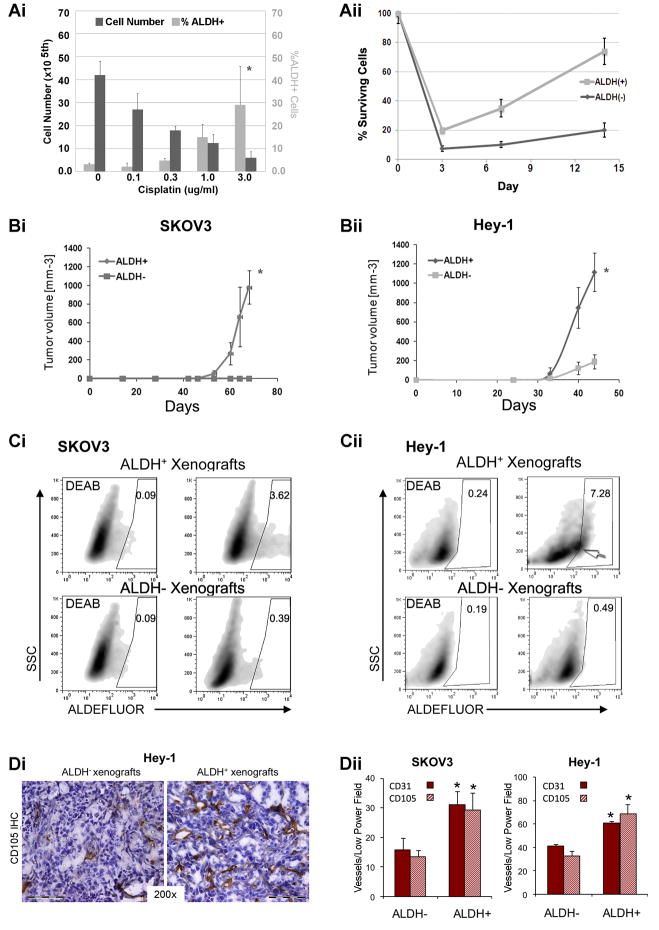
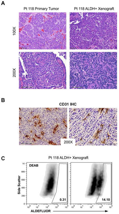
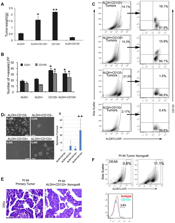
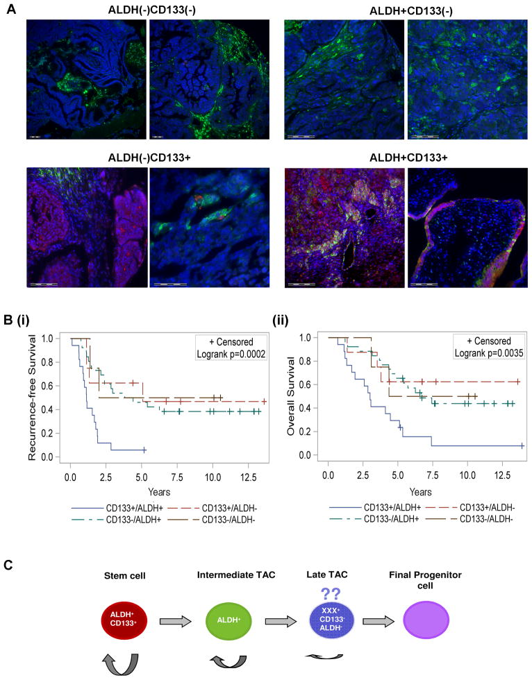
Markers that reliably identify cancer stem cells (CSC) in ovarian cancer could assist prognosis and improve strategies for therapy. CD133 is a reported marker of ovarian CSC. Aldehyde dehydrogenase (ALDH) activity is a reported CSC marker in several solid tumors, but it has not been studied in ovarian CSC. Here we report that dual positivity of CD133 and ALDH defines a compelling marker set in ovarian CSC. All human ovarian tumors and cell lines displayed ALDH activity. ALDH(+) cells isolated from ovarian cancer cell lines were chemoresistant and preferentially grew tumors, compared with ALDH(-) cells, validating ALDH as a marker of ovarian CSC in cell lines. Notably, as few as 1,000 ALDH(+) cells isolated directly from CD133(-) human ovarian tumors were sufficient to generate tumors in immunocompromised mice, whereas 50,000 ALDH(-) cells were unable to initiate tumors. Using ALDH in combination with CD133 to analyze ovarian cancer cell lines, we observed even greater growth in the ALDH(+)CD133(+) cells compared with ALDH(+)CD133(-) cells, suggesting a further enrichment of ovarian CSC in ALDH(+)CD133(+) cells. Strikingly, as few as 11 ALDH(+)CD133(+) cells isolated directly from human tumors were sufficient to initiate tumors in mice. Like other CSC, ovarian CSC exhibited increased angiogenic capacity compared with bulk tumor cells. Finally, the presence of ALDH(+)CD133(+) cells in debulked primary tumor specimens correlated with reduced disease-free and overall survival in ovarian cancer patients. Taken together, our findings define ALDH and CD133 as a functionally significant set of markers to identify ovarian CSCs.


Conflict of interest statement

Conflict of Interest

The authors have no conflicts of interest to report

Figures

Figure 1
Figure 1. In vitro and in vivo outgrowth of ALDH+ cells human ovarian cancer cell lines
(Ai) Absolute cell number and %ALDH+ SKOV3 cells following treatment with indicated concentrations of Cisplatin. (Aii) Percent viable FACS sorted ALDH and ALDH+ cells prior to and 3, 7, or 14 days following treatment with 1.5 μg/ml Cisplatin. (Bi and ii) Tumor growth curves of 100 FACS isolated ALDH+ and ALDH SKOV3 and Hey1 cells. (Ci and ii) ALDEFLUOR staining of ALDH+ and ALDH SKOV3 and Hey1 tumor xenografts with DEAB controls. (D) CD105 IHC from Hey-1 ALDH and ALDH+ xenografts tumors. (E) Quantification of CD31+ and CD105+ microvascular density in the indicated tumor xenografts. Scale bar indicates 100μm. All data is representative of at least 2 independent experiments (n=5 tumors/experiment). * Indicates p<0.01 versus controls.
Figure 2
Figure 2. In vivo tumor generation from FACS isolated ALDH+ primary human epithelial ovarian tumor cells
(A) Histology of primary tumor and tumor xenograft generated from ALDH+ cells isolated from Pt118’s tumor. (B) CD31 IHC analysis of primary ovarian ALDH+ cell derived tumor xenografts. (C) FACS analysis demonstrating DEAB control (left) and ALDEFLUOR activity (right) from Pt 118 ALDH+ cell tumor xenograft second passage.
Figure 3
Figure 3. Characterization of ALDH+CD133+ cells as CSC in ovarian tumor cell lines and primary human epithelial ovarian tumors
(A) Tumor weights from 1000 cells of the indicated FACS isolated A2780 tumor cell populations (n=5/group). (B) Quantification of CD31+ and CD105+ microvascular density (vessels/low power field) of the indicated A2780 cell line populations. (C) FACS analysis of indicated A2780 xenografts for expression ALDH, and CD133 in ALDH+ gate. (Di) Spheres generated from FACS isolated ALDH−/+CD133+/− cells from a primary ovarian tumor sample. Cell numbers plated at the initiation of the sphere assay are indicated. Scale bar (lower left) indicates 100μm. (ii) Average sphere formation in the indicated cell population from 8 different patients. (E) Histology of Pt94 primary tumor and tumor xenograft generated from ALDH+CD133+ isolated from Pt94. (F) FACS analysis of Pt 94 tumor, left panel demonstrates DEAB control, middle demonstrates ALDEFLUOR activity in Pt 94 xenograft, right demonstrates histogram for CD133 with isotype control. *indicates p<0.05 compared to ALDH+CD133 and ALDHCD133 groups, ** indicates p<0.05 versus all other groups.
Figure 4
Figure 4. ALDH+CD133+ cells identify poor risk tumors
(A) Representative Immunofluorescent classification of ovarian tumors as ALDH+/−CD133+/−; ALDH is stained in green and CD133 in red. Scale bars indicate 100 micrometers. (Bi and ii) Kaplan-Meier analysis of overall and recurrence free survival in 56 patients whose tumors were scored as either ALDHCD133 (n=4), ALDH+CD133 (n=26), ALDH−CD133+ (n=8), or ALDH+CD133+ (n=18). See methods for scoring methodology. (C) Proposed model for ovarian cancer stem cell differentiation. We speculate an ALDH+CD133+ CSC gives rise to an ALDH+CD133 transient amplifying cell (TAC). This cell subsequently gives rise to an ALDHCD133 late TAC. The ability to self replicate is indicated by semi-circular arrow, with cells on the left having greatest self-renewal capacity and cells to the right having minimal self-renewal capacity.

References

    1. Quintana E, Shackleton M, Sabel MS, Fullen DR, Johnson TM, Morrison SJ, et al. Efficient tumour formation by single human melanoma cells. Nature. 2008;456:593–8. - PMC - PubMed
    1. Boman BM, Wicha MS. Cancer stem cells: a step toward the cure. Journal of Clinical Oncology. 2008;26:2795–9. - PubMed
    1. Zhang S, Balch C, Chan MW, Lai H-C, Matei D, Schilder JM, et al. Identification and characterization of ovarian cancer-initiating cells from primary human tumors. Cancer Research. 2008;68:4311–20. - PMC - PubMed
    1. Baba T, Convery PA, Matsumura N, Whitaker RS, Kondoh E, Perry T, et al. Epigenetic regulation of CD133 and tumorigenicity of CD133+ ovarian cancer cells. Oncogene. 2009;28:209–18. - PubMed
    1. Curley MD, Therrien VA, CL C. CD133 Expression Defines a Tumor Initiating Cell Population in Primary Human Ovarian Cancer. Stem Cells. 2009 - PubMed

Publication types

MeSH terms