. 2011 Apr;23(4):1435-48.
doi: 10.1105/tpc.110.082768. Epub 2011 Apr 15.

The Arabidopsis thaliana checkpoint kinase WEE1 protects against premature vascular differentiation during replication stress

Affiliations

The Arabidopsis thaliana checkpoint kinase WEE1 protects against premature vascular differentiation during replication stress

Toon Cools et al. Plant Cell. 2011 Apr.

Abstract

A sessile lifestyle forces plants to respond promptly to factors that affect their genomic integrity. Therefore, plants have developed checkpoint mechanisms to arrest cell cycle progression upon the occurrence of DNA stress, allowing the DNA to be repaired before onset of division. Previously, the WEE1 kinase had been demonstrated to be essential for delaying progression through the cell cycle in the presence of replication-inhibitory drugs, such as hydroxyurea. To understand the severe growth arrest of WEE1-deficient plants treated with hydroxyurea, a transcriptomics analysis was performed, indicating prolonged S-phase duration. A role for WEE1 during S phase was substantiated by its specific accumulation in replicating nuclei that suffered from DNA stress. Besides an extended replication phase, WEE1 knockout plants accumulated dead cells that were associated with premature vascular differentiation. Correspondingly, plants without functional WEE1 ectopically expressed the vascular differentiation marker VND7, and their vascular development was aberrant. We conclude that the growth arrest of WEE1-deficient plants is due to an extended cell cycle duration in combination with a premature onset of vascular cell differentiation. The latter implies that the plant WEE1 kinase acquired an indirect developmental function that is important for meristem maintenance upon replication stress.


Figures

Figure 1.
Figure 1.
Lack of Hypersensitivity to BLM of WEE1KO Plants. Plants of the indicated genotypes were germinated on 0.6 μg/mL BLM, after which root length was measured at the indicated time points. Data are means ± se (n > 21)
Figure 2.
Figure 2.
Clustering of Microarray Data of HU-Treated Col-0 and WEE1KO. Genes with significantly altered transcription levels upon HU treatment were clustered into seven different groups based on their expression levels at different time points in mutant and wild-type root tips. Each panel shows the mean normalized expression values of all the genes within the cluster for Col-0 (blue) and WEE1KO (red) after 0, 5, and 24 h on HU. The number of genes that each cluster contains is given on the right.
Figure 3.
Figure 3.
Time-Course Analysis of Cell Cycle Genes in HU-Synchronized Col-0 and WEE1KO Root Tips. Transcript levels were measured by nCounter analysis. Fold induction levels of the different transcripts are presented for Col-0 (blue) and WEE1KO (red). EMB2386, PAC1, and RPS26C were used as reference genes. Transcript levels were rescaled to the level in Col-0 at 0 h (=1). Time after treatment with HU is given on the horizontal axis. Data are means ± se. (A) CYCA3;1. (B) Histone H4. (C) Histone H2B. (D) Histone H1. (E) CYCA2;1. (F) CYCB1;2. (G) CYCB1;1. (H) BRCA1.
Figure 4.
Figure 4.
Stabilization of WEE1 during S Phase upon Replication Stress. (A) Transcript analysis of CYCA3;1 (blue) and WEE1 (red) in HU-synchronized Col-0 root tips. Data are means ± se. (B) Daily growth of Col-0 (blue), WEE1KO (red), and PWEE1:GFP-WEE1 (green) roots after transfer to 1 mM HU. Growth was followed until 4 d after transfer. Data are means ± se (n > 37). (C) Confocal microscopy of the PWEE1:GFP-WEE1 line transferred for 24 h to 0 mM HU (top) and 1 mM HU (bottom). Epidermis-localized green signal at 0 mM HU is due to background signal. Nuclear GFP-WEE1 signal in the vascular cells was seen after treatment with 1 mM HU. Bar = 0.1 mm. (D) Flow cytometry profile of unsorted and untreated PWEE1:GFP-WEE1 root tips. 2C, 4C, and S indicate G1, G2, or S-phase nuclei, respectively. (E) Flow cytometry profile of unsorted (red) and sorted GFP-WEE1 (blue) root tips treated for 24 h with 10 mM HU.
Figure 5.
Figure 5.
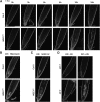
Vascular Cell Death Phenotype of WEE1KO roots upon Replication Stress. (A) Time course of Col-0 and WEE1KO root tips transferred to HU and stained with confocal microscopy. Dead cells are stained with PI. Time after transfer to HU medium is indicated above the root tips. (B) PI staining of Col-0 and WEE1KO after treatment with 0.6 μg/mL BLM for 24 h. (C) PI staining of Col-0 and WEE1KO grown for 24 h on 5 mM HU. (D) PI staining of atm-1 and atr-2 transferred either for 24 h to control medium or 1 mM HU.
Figure 6.
Figure 6.
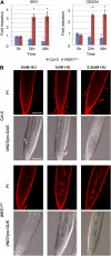
Premature Vascular Differentiation Induced by Replication Stress in WEE1KO Plants. (A) Tracheary element differentiation genes are induced in WEE1KO root tips upon replication stress. qRT-PCR transcript analysis of IRX1 and CESA4 in Col-0 and WEE1KO 0, 24, and 48 h after HU treatment. EMB2386, PAC1, and RPS26C were used as reference genes. The transcript levels were rescaled to the level in Col-0 at 0 h (=1). Asterisks indicate significantly altered transcription levels compared with the 0-h time point (*P value < 0.05; **P value <0.001). Data are means ± se (n = 4). (B) Confocal microscopy of Col-0 and WEE1KO plants after PI staining to detect cell death. Col-0 and WEE1KO harboring a VND7pro:GUS construct were stained with GUS. Plants were treated either with 0 mM (left), 1 mM HU (center), or 2.5 mM HU (right) for 24 h. Bars = 0.1 mm.
Figure 7.
Figure 7.
Reduced Vascular Cell Death in WEE1KO by BA. PI staining to detect cell death in WEE1KO plants treated for 24 h (top) or 48 h (bottom) with 100 nM BA (left), 1 mM HU (center), or 1 mM HU and 100 nM BA (right). To show the reproducibility of the experiment, several treated root tips are shown in Supplemental Figure 4 online. Bar = 0.1 mm.
Figure 8.
Figure 8.
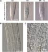
Aberrant Vascular Differentiation in WEE1KO Plants Caused by HU Treatment. (A) GUS staining of VND7pro:GUS (WEE1KO) after 24 h treatment with 100 nM BA (left), 2.5 mM HU (center), or a combination of both BA and HU (right). Bar = 0.1 mm. (B) Microscopy analysis of vascular development of Col-0 treated for 4 d with 2.5 mM HU. Bar = 0.05 mm. (C) Microscopy analysis of aberrant vascular development of WEE1KO plants treated for 4 d with 2.5 mM HU. Bar = 0.05 mm.

References

    1. Alvino G.M., Collingwood D., Murphy J.M., Delrow J., Brewer B.J., Raghuraman M.K. (2007). Replication in hydroxyurea: It's a matter of time. Mol. Cell. Biol. 27: 6396–6406 - PMC - PubMed
    1. Beeckman T., Engler G. (1994). An easy technique for the clearing of histochemically stained plant tissue. Plant Mol. Biol. Rep. 12: 37–42
    1. Birnbaum K., Shasha D.E., Wang J.Y., Jung J.W., Lambert G.M., Galbraith D.W., Benfey P.N. (2003). A gene expression map of the Arabidopsis root. Science 302: 1956–1960 - PubMed
    1. Boudolf V., Barrôco R., de Almeida Engler J., Verkest A., Beeckman T., Naudts M., Inzé D., De Veylder L. (2004). B1-type cyclin-dependent kinases are essential for the formation of stomatal complexes in Arabidopsis thaliana. Plant Cell 16: 945–955 - PMC - PubMed
    1. Boudolf V., Inzé D., De Veylder L. (2006). What if higher plants lack a CDC25 phosphatase? Trends Plant Sci. 11: 474–479 - PubMed

Publication types

MeSH terms