Review
. 2011 Jun;17(3):321-36.
doi: 10.1177/1073858410384740. Epub 2011 Apr 15.

AKAP signaling complexes in regulation of excitatory synaptic plasticity

Affiliations
Review

AKAP signaling complexes in regulation of excitatory synaptic plasticity

Jennifer L Sanderson et al. Neuroscientist. 2011 Jun.

Abstract

Plasticity at excitatory glutamatergic synapses in the central nervous system is believed to be critical for neuronal circuits to process and encode information, allowing animals to perform complex behaviors such as learning and memory. In addition, alterations in synaptic plasticity are associated with human diseases, including Alzheimer disease, epilepsy, chronic pain, drug addiction, and schizophrenia. Long-term potentiation (LTP) and depression (LTD) in the hippocampal region of the brain are two forms of synaptic plasticity that increase or decrease, respectively, the strength of synaptic transmission by postsynaptic AMPA-type glutamate receptors. Both LTP and LTD are induced by activation of NMDA-type glutamate receptors but differ in the level and duration of Ca(2+) influx through the NMDA receptor and the subsequent engagement of downstream signaling by protein kinases, including PKA, PKC, and CaMKII, and phosphatases, including PP1 and calcineurin-PP2B (CaN). This review addresses the important emerging roles of the A-kinase anchoring protein family of scaffold proteins in regulating localization of PKA and other kinases and phosphatases to postsynaptic multiprotein complexes that control NMDA and AMPA receptor function during LTP and LTD.


Figures
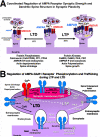
Figure 1
Figure 1. Mechanisms of Postsynaptic Plasticity
A) Regulation of NMDAR-induced LTP and LTD in dendritic spines. LTD induces spine shrinkage, AMPAR dephosphorylation, and AMPAR removal from spines through protein phosphatase activity. LTP induces spine growth, AMPAR phosphorylation, and AMPAR recruitment to spines through protein kinase activity. B) Regulation of AMPAR-GluR1 subunit phosphorylation and trafficking to and from synapses during LTP and LTD. These trafficking events may be similar for both GluR1/2 and GluR1/1 receptors. During LTP, AMPARs may be secreted to the extrasynaptic membrane directly in spines or to regions of dendrite shafts near spines (not depicted). Likewise, during LTD AMPAR endocytosis may occur both within spines and on dendrite shafts (not depicted).
Figure 2
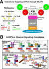
Figure 2. Subcellular targeting of PKA and other signaling proteins by AKAPs
A) Left, schematic representation of different converging signaling pathways leading to adenylyl cyclase production of cAMP and activation of the PKA holoenzyme. Right, schematic regulation of the AKAP-anchored PKA holoenzyme. B) Examples of AKAP organized glutamate receptor and ion channel signaling complexes. Top, AKAP79/150 organized signaling complexes. Bottom, Yotiao, MAP2, and AKAP15/18 organized signaling complexes.
Figure 3
Figure 3. The AKAP79/150-MAGUK postsynaptic signaling complex in regulation of AMPA receptor phosphorylation during synaptic plasticity
A) AKAP79/150 can be linked to both AMPA and NMDA receptors through MAGUK scaffolding proteins. Influx of Ca2+ through the NMDAR activates CaN, PKC, and PKA activity to regulate postsynaptic substrate phosphorylation including AMPAR-GluR1. B) Regulation of the AMPAR endocytosis and translocation of AKAP79/150 from the PSD during LTD. Ca2+ influx through the NMDAR activates AKAP-anchored CaN, resulting in dephosphorylation of the GluR1-Ser845 and AMPAR endocytosis. Activation of CaN along with PLC also promotes depolymerization of spine actin to promote subsequent translocation of AKAP-PKA complexes away from the postsynaptic membrane in dendritic spines. This delayed movement of AKAP79/150-PKA away from spines may prevent re-phosphorylation and recycling of AMPARs during LTD.
Figure 4
Figure 4. Domain Organization of the AKAP79/150 Signaling Scaffold
Amino acid numbering is given for human AKAP79 from 1 at the N-terminus to 427 at the C-terminus. The locations of the various mapped binding sites and the indicated binding partners are shown. See the text for more details. The repetitive sequence unique to rodent AKAP150 which gives it a highe molecular weight (150 kDa vs. 79kDa for human) is inserted near residue 315 between the MAGUK binding and the CaN anchoring domains.
Figure 5
Figure 5. AKAP79/150 is co-localized with PKA and CaN in hippocampal neuron dendrites
A) Immunofluorescent staining for rat AKAP150 (red) and the PKA-RIIβ regulatorysubunit (green) shows punctate co-localization (yellow in merge panels) along hippocampal neurons dendrites including in dendritic spines. Right hand panels are magnifications of dendrites. Prominent localization of PKA-RIIβ independent of AKAP150 is also seen in the cell body and the interior of dendrite shafts where it may bind other AKAPs including MAP2. B) Immunofluorescent staining for AKAP150 (red) and CaNB regulatory subunit (green) shows punctate co-localization (yellow in merge panels) along hippocampal neurons dendrites including in dendritic spines. Right hand panels are magnifications of dendrites. Prominent localization of CaNB independent of AKAP150 is also seen in the cell body and in likely axonal/presynaptic punctate along dendrites that are in most cases closely opposed to the sites of dendritic co-localization (yellow) between AKAP150 and CaN. See (Gomez Alam Smith Horne and Dell'Acqua 2002) for the original report of these findings.
Figure 6
Figure 6. AKAP79/150 PKA anchoring controls dendritic spine localization of PKA
A) Co-expression of AKAP79WT-YFP (green) and PKA-RIIα-CFP (blue) in rat hippocampal neurons leads to co-localization (turquoise in merge panels) of PKA-RII in spines with the AKAP79. B) However, co-expression of the 1-360 truncation mutant AKAP79ΔPKA-YFP (green) with PKA-RIIα-CFP (blue) fails to target PKA-RII to spine leading to its retention in the interior of dendrite shafts likely through anchoring to MAP2. Right hand panels are magnifications of dendrites. See (Smith Gibson and Dell'Acqua 2006) for the original report of these findings.
Figure 7
Figure 7. AKAP79/150 postsynaptic co-localization with PSD-95 is disrupted following chemical induction of LTD in hippocampal neurons
A) Immunofluorescent staining for rat AKAP150 (red) and the PSD-95 (green) shows punctate co-localization (yellow in merge panels) along hippocampal neurons dendrites including in dendritic spines in control neurons. B) AKAP150 staining declusters and redistributes away from spines and PSD-95 puncta toward dendrite shafts and the soma 30 minutes following a chemical LTD induction stimulus (25 μM NMDA, 3 minutes). Right hand panels are magnifications of dendrites. See (Smith Gibson and Dell'Acqua 2006) for the original report of these findings.
Figure 8
Figure 8. FRET imaging reveals redistribution of AKAP79-PKA anchoring from dendritic spines to dendrite shafts following chemical induction of LTD in living hippocampal neurons
A) Diagram showing design of the FRET imaging approach used to detect changes in the localization of AKAP79-PKA anchoring within hippocampal neurons. B) Corrected images of sensitized FRET emission from PKA-RIIα-YFP at 535 nM upon excitation of AKAP79-CFP at 436 nm (pseudocolor scale: blue=no FRET to red=high FRET) for dendrites from control untreated neurons or 15 minutes after chemical LTD induction (25 μM NMDA, 3 minutes). See (Oliveria Gomez and Dell'Acqua 2003; Smith Gibson and Dell'Acqua 2006) for the original reports using this FRET imaging method.
Figure 9
Figure 9. Deficits in postsynaptic PKA localization and hippocampal synaptic plasticity in AKAP150 mutant mice
A) Immunohistochemical (IHC) staining for PKA-RIIα shows decreased PKA localization in CA1 dendritic regions and increased PKA localization in the cell body layer (red arrows) in AKAP150 knockout (KO) and D36 mice compared to WT. B) PKA-RIIα immunoblotting shows decreased PKA levels in synaptic membrane pellet (P) fractions and increased PKA levels in cytoplasmic supernatant (S) fractions in AKAP150 KO and D36 mice compared to WT. AKAP150 immunoblotting confirms lack of AKAP150 expression in KO mice and normal expression and distribution in D36 mice compared to WT. C) AKAP150 D36 mice but not AKAP150 KO mice exhibit reduced CA1 hippocampal LTP at 7-12 weeks of age and D) LTD at ~2 weeks (10-14 days) of age compared to WT littermate mice. Shown are plots of the average field EPSP initial slope measured 55-60 minutes after induction of LTD (1Hz, 15 min) or LTP (100Hz, 1 sec) as a % of the baseline initial slope (100%) measure 5 minutes prior to induction. WT and KO mice show ~45% potentiation with LTP and ~35% depression with LTD, while D36 mice show only ~15% potentiation with LTP and ~10% depression with LTD. *p<0.05 by ANOVA. Reproduced and adapted from (Weisenhaus and others 2010).

References

    1. Adesnik H, Nicoll RA. Conservation of glutamate receptor 2-containing AMPA receptors during long-term potentiation. J Neurosci. 2007;27(17):4598–602. - PMC - PubMed
    1. Bal M, Zhang J, Hernandez CC, Zaika O, Shapiro MS. Ca2+/calmodulin disrupts AKAP79/150 interactions with KCNQ (M-Type) K+ channels. J Neurosci. 2010;30(6):2311–23. - PMC - PubMed
    1. Banke TG, Bowie D, Lee H, Huganir RL, Schousboe A, Traynelis SF. Control of GluR1 AMPA receptor function by cAMP-dependent protein kinase. J Neurosci. 2000;20(1):89–102. - PMC - PubMed
    1. Barria A, Derkach V, Soderling T. Identification of the Ca2+/calmodulin-dependent protein kinase II regulatory phosphorylation site in the alpha-amino-3-hydroxyl-5-methyl-4-isoxazole-propionate-type glutamate receptor. J Biol Chem. 1997;272(52):32727–30. - PubMed
    1. Barria A, Muller D, Derkach V, Griffith LC, Soderling TR. Regulatory phosphorylation of AMPA-type glutamate receptors by CaM-KII during long-term potentiation. Science. 1997;276(5321):2042–5. - PubMed

Publication types

Substances

LinkOut - more resources