

In silico and in vivo investigations of proteins of a minimized eukaryotic cytoplasm
- PMID: 21498883
- PMCID: PMC3101018
- DOI: 10.1093/gbe/evr031
In silico and in vivo investigations of proteins of a minimized eukaryotic cytoplasm
Abstract
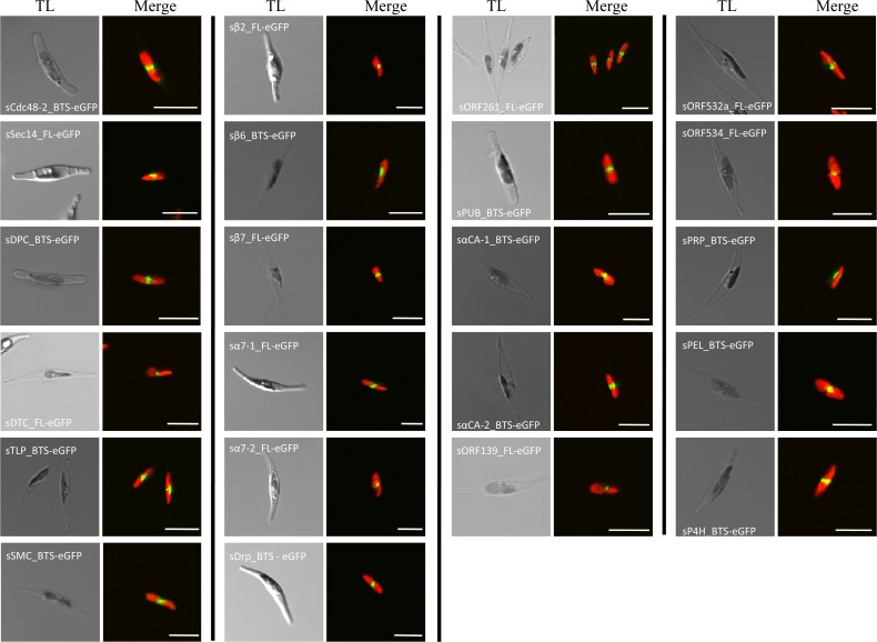
Algae with secondary plastids such as diatoms maintain two different eukaryotic cytoplasms. One of them, the so-called periplastidal compartment (PPC), is the naturally minimized cytoplasm of a eukaryotic endosymbiont. In order to investigate the protein composition of the PPC of diatoms, we applied knowledge of the targeting signals of PPC-directed proteins in searches of the genome data for proteins acting in the PPC and proved their in vivo localization via expressing green fluorescent protein (GFP) fusions. Our investigation increased the knowledge of the protein content of the PPC approximately 3-fold and thereby indicated that this narrow compartment was functionally reduced to some important cellular functions with nearly no housekeeping biochemical pathways.
Figures

Similar articles
-
The periplastidal compartment: a naturally minimized eukaryotic cytoplasm.Curr Opin Microbiol. 2014 Dec;22:88-93. doi: 10.1016/j.mib.2014.09.017. Curr Opin Microbiol. 2014. PMID: 25460801 Review.
-
Characterization of periplastidal compartment-targeting signals in chlorarachniophytes.Mol Biol Evol. 2010 Jul;27(7):1538-45. doi: 10.1093/molbev/msq038. Epub 2010 Feb 4. Mol Biol Evol. 2010. PMID: 20133351
-
ERAD-derived preprotein transport across the second outermost plastid membrane of diatoms.Mol Biol Evol. 2009 Aug;26(8):1781-90. doi: 10.1093/molbev/msp079. Epub 2009 Apr 17. Mol Biol Evol. 2009. PMID: 19377060
-
Nucleus-to-nucleus gene transfer and protein retargeting into a remnant cytoplasm of cryptophytes and diatoms.Mol Biol Evol. 2006 Dec;23(12):2413-22. doi: 10.1093/molbev/msl113. Epub 2006 Sep 13. Mol Biol Evol. 2006. PMID: 16971693
-
Transit peptide diversity and divergence: A global analysis of plastid targeting signals.Bioessays. 2007 Oct;29(10):1048-58. doi: 10.1002/bies.20638. Bioessays. 2007. PMID: 17876808 Review.
Cited by
-
An Enigmatic Stramenopile Sheds Light on Early Evolution in Ochrophyta Plastid Organellogenesis.Mol Biol Evol. 2022 Apr 11;39(4):msac065. doi: 10.1093/molbev/msac065. Mol Biol Evol. 2022. PMID: 35348760 Free PMC article.
-
Proteomic amino-termini profiling reveals targeting information for protein import into complex plastids.PLoS One. 2013 Sep 16;8(9):e74483. doi: 10.1371/journal.pone.0074483. eCollection 2013. PLoS One. 2013. PMID: 24066144 Free PMC article.
-
Plastid proteome prediction for diatoms and other algae with secondary plastids of the red lineage.Plant J. 2015 Feb;81(3):519-28. doi: 10.1111/tpj.12734. Epub 2015 Jan 6. Plant J. 2015. PMID: 25438865 Free PMC article.
-
In vivo localization of iron starvation induced proteins under variable iron supplementation regimes in Phaeodactylum tricornutum.Plant Direct. 2022 Dec 26;6(12):e472. doi: 10.1002/pld3.472. eCollection 2022 Dec. Plant Direct. 2022. PMID: 36582220 Free PMC article.
-
Genome and methylome of the oleaginous diatom Cyclotella cryptica reveal genetic flexibility toward a high lipid phenotype.Biotechnol Biofuels. 2016 Nov 25;9:258. doi: 10.1186/s13068-016-0670-3. eCollection 2016. Biotechnol Biofuels. 2016. PMID: 27933100 Free PMC article.
References
-
- Allen MD, Buchberger A, Bycroft M. The PUB domain functions as a p97 binding module in human peptide N-glycanase. J Biol Chem. 2006;281:25502–25508. - PubMed
-
- Archibald JM. The puzzle of plastid evolution. Curr Biol. 2009;19:R81–R88. - PubMed
-
- Beskow A, et al. A conserved unfoldase activity for the p97 AAA-ATPase in proteasomal degradation. J Mol Biol. 2009;394:732–746. - PubMed
Publication types
MeSH terms
Substances
LinkOut - more resources
Full Text Sources
