. 2011 Jun 17;286(24):21755-66.
doi: 10.1074/jbc.M111.218669. Epub 2011 Apr 17.

Drosophila melanogaster myosin-18 represents a highly divergent motor with actin tethering properties

Affiliations

Drosophila melanogaster myosin-18 represents a highly divergent motor with actin tethering properties

Stephanie Guzik-Lendrum et al. J Biol Chem. .

Abstract

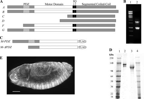
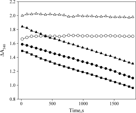
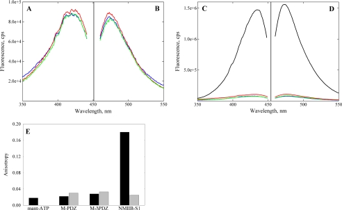
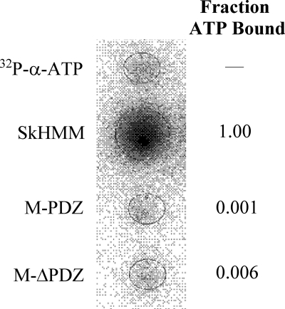
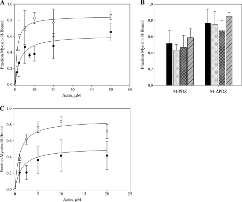
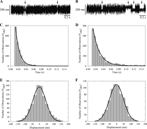
The gene encoding Drosophila myosin-18 is complex and can potentially yield six alternatively spliced mRNAs. One of the major features of this myosin is an N-terminal PDZ domain that is included in some of the predicted alternatively spliced products. To explore the biochemical properties of this protein, we engineered two minimal motor domain (MMD)-like constructs, one that contains the N-terminal PDZ (myosin-18 M-PDZ) domain and one that does not (myosin-18 M-ΔPDZ). These two constructs were expressed in the baculovirus/Sf9 system. The results suggest that Drosophila myosin-18 is highly divergent from most other myosins in the superfamily. Neither of the MMD constructs had an actin-activated MgATPase activity, nor did they even bind ATP. Both myosin-18 M-PDZ and M-ΔPDZ proteins bound to actin with K(d) values of 2.61 and 1.04 μM, respectively, but only about 50-75% of the protein bound to actin even at high actin concentrations. Unbound proteins from these actin binding assays reiterated the 60% saturation maximum, suggesting an equilibrium between actin-binding and non-actin-binding conformations of Drosophila myosin-18 in vitro. Neither the binding affinity nor the substoichiometric binding was significantly affected by ATP. Optical trapping of single molecules in three-bead assays showed short lived interactions of the myosin-18 motors with actin filaments. Combined, these data suggest that this highly divergent motor may function as an actin tethering protein.


Figures

FIGURE 1.
FIGURE 1.
Sequence analysis of PDZ and ΔPDZ isoforms of Drosophila myosin-18. A, sequence analysis of 17 exons in the Drosophila myosin-18 gene (CG31045) was performed using the ClustalW multiple alignment algorithm. The domain analysis using Simple Modular Architecture Research Tool (SMART) at European Molecular Biology Laboratory was aligned with exons to illustrate six alternatively spliced isoforms (A–G) varying at the N terminus. B, lane 1 is molecular weight standards. Lane 2, 5′ RACE analysis of whole fly mRNA using nested PCR to amplify PDZ and ΔPDZ isoforms yielded two bands, one at 1.7 kb corresponding to PDZ-containing isoforms and one at 0.9 kb corresponding to the ΔPDZ isoform. C, expression constructs of minimal motors of Drosophila myosin-18 for baculoviral expression in Sf9 cells truncate the sequence at Leu1319 for M-PDZ and Leu1082 for M-ΔPDZ, the residues corresponding to Dictyostelium myosin-2 Arg761, followed by a C-terminal FLAG tag for purification. D, M-PDZ and M-ΔPDZ expressed proteins purified using a FLAG affinity tag and fractionated by 4–20% Tris-glycine SDS-PAGE. Lane 1, molecular weight markers; lane 2, M-PDZ at 140 kDa; lane 3, M-ΔPDZ at 116 kDa: lane 4, molecular weight markers. E, representative Drosophila embryo (stage 13) stained with a polyclonal antibody at a 1:1,000 dilution in blocking buffer (scale bar, 50 μm). The primary antibody was raised in rabbit against an uninterrupted segment of the coiled coil region of myosin-18 that is shared between all six potential isoforms of the protein. Visualization of the localization pattern used Alexa Fluor 488 goat anti-rabbit secondary antibody at a 1:5,000 dilution in blocking buffer. Using confocal microscopy with a 20×, 0.75 numerical aperture oil immersion objective and a section depth of 1.2 μm, the localization of myosin-18 was ubiquitous throughout all embryonic tissues as seen in this projection of 17 confocal sections. Such ubiquitous staining was seen throughout all observed embryonic developmental stages.
FIGURE 2.
FIGURE 2.
Drosophila myosin-18 lacks actin-activated MgATPase activity. M-PDZ and M-ΔPDZ were assayed for actin-activated MgATPase activity in an NADH-coupled assay. Representative traces of data from M-PDZ (○) and M-ΔPDZ (△) motors in the absence of actin show no detectable hydrolysis of ATP. In the presence of 45 μm F-actin and 2 mm ATP, M-PDZ (●) and M-ΔPDZ (▴) show no detectable difference in the rate of change of A340 from the ATP hydrolysis rate of actin alone (■) at the same concentration. Experiments were conducted at 25 °C in a buffer containing 50 mm KCl, 10 mm MOPS (pH 7.2), 2 mm MgCl2, 0.15 mm EGTA, 2 mm ATP, 40 units/ml lactate dehydrogenase, 200 units/ml pyruvate kinase, 1 mm phosphoenolpyruvate, and 200 μm NADH. The concentration of myosin-18 fragments in each assay was 0.5 μm.
FIGURE 3.
FIGURE 3.
Excitation and emission spectra of deac-amino nucleotides in presence and absence of myosin-18. Excitation (A) and emission (B) spectra of deac-amino-ATP in binding assays using expressed M-PDZ (red) and M-ΔPDZ (green) in comparison with base-line fluorescence of the deac-amino moiety (blue) are shown. Fluorometric analysis of fluorescence signals from the deac moiety exhibited peak excitation at 430 nm and emission at 470 nm at 20 °C. Assays used 1 μm M-PDZ or M-ΔPDZ and 0.5 μm deac-amino-ATP in 0.5 m KCl, 10 mm MOPS (pH 7.2), 3 mm MgCl2, 1 mm EGTA, and 1 mm DTT. The proteins were spun for 10 min at 100,000 × g in a Beckman TLA-100 rotor at 4 °C just prior to the assay to remove aggregates. C and D, excitation and emission spectra of deac-amino-ATP assays repeated and contrasted with 1 μm mouse myosin-5-S1–6IQ (black). Myosin-5-S1–6IQ was analyzed in a similar buffer as above but containing 25 mm KCl. Note the difference in the scale values for the fluorescence intensities in the two experiments. E, steady-state fluorescence anisotropy values comparing mant-ATP at 0.5 μm (black) alone in solution with mant-ATP in the presence of Drosophila myosin-18 M-PDZ and -ΔPDZ proteins at 1.96 and 2.63 μm, respectively. Experiments were performed in buffer containing 0. 5 m KCl, 10 mm MOPS (pH 7.2), 0.1 mm EGTA, 3 mm NaN3, and 1 mm DTT. Controls using 6.1 μm NMIIB S1 and further controls for viscosity effects by adding 2 mm unlabeled ATP (gray) are also shown. cps, counts/s.
FIGURE 4.
FIGURE 4.
Binding of [α-32P]ATP to myosin via filter binding assay. Rabbit SkHMM, M-PDZ, and M-ΔPDZ at 1 μm were incubated with 20 μm [α-32P]ATP (1.3 × 10¹⁵ cpm/mol) for 60 s and blotted onto a nitrocellulose membrane pre-equilibrated with buffer containing 0.25 m KCl, 10 mm MOPS (pH 7.2), 0.1 mm EGTA, 3 mm NaN3, and 1 mm DTT under vacuum. Blots of 20 μm [α-32P]ATP (1.3 × 10¹⁵ cpm/mol) were used as controls. Following a rinse with excess equilibration buffer, the membrane was dried and exposed to Fujifilm BAS-MS phosphorimaging screens for 1 h. The fraction of [α-32P]ATP bound to M-PDZ and M-ΔPDZ was calculated in relation to SkHMM after correcting for the amount of nucleic acid that binds nitrocellulose in the absence of protein.
FIGURE 5.
FIGURE 5.
CD spectrum of temperature-dependent unfolding of Drosophila myosin-18 proteins. Data were collected at 222 nm over a range of temperatures determined at Tm = 45.4 ± 0.1 °C for M-PDZ (●) and Tm = 46.3 ± 0.3 °C M-ΔPDZ (○). Both proteins were dialyzed into buffer containing 0.5 m KCl, 10 mm KH2PO4 (pH 7.2), 0.1 mm EGTA, 3 mm NaN3, and 1 mm DTT prior to the assay. The final concentration of M-PDZ used in the assay was 1.78 μm, and that of M-ΔPDZ was 2.85 μm. mdeg, millidegrees.
FIGURE 6.
FIGURE 6.
Binding of myosin-18 isoforms to actin. A, Drosophila myosin-18 motor constructs at a final concentration of 1 μm in buffer containing 0.1 mm KCl, 20 mm MOPS (pH 7.0), 5 mm MgCl2, 0.05 mm EGTA, 1 mm NaN3, and 1 mm DTT were incubated for 10 min at room temperature with increasing concentrations of phalloidin-stabilized F-actin in the absence of ATP. Reactions were sedimented at 100,000 × g for 15 min. Pellets and supernatants were separated by SDS-PAGE, and gels were stained and imaged with Coomassie Blue. Fractions of motor pelleted for each reaction were calculated by densitometry, correcting for the amount of motor that pellets itself in the absence of actin, typically in the range of 10%, for each preparation of protein. The fraction of myosin-18 motor bound was plotted against the concentration of actin introduced in each reaction. Data collected from M-PDZ (●) defined a Kd of 2.6 ± 0.2 μm and saturation at 58.2 ± 11.0%. Data from M-ΔPDZ (○) resulted in a Kd of 1.0 ± 0.2 μm and saturation at 83.2 ± 5.4%. The data represent multiple rounds of binding experiments using at least six different preparations of purified motor constructs for data collection. Error bars represent standard deviation of the mean (mean ± standard deviation). B, comparison of variations in the cosedimentation assay using M-PDZ and M-ΔPDZ. Cosedimentations at 20 μm actin in A (■) were compared with variations of the parameters within the cosedimentation assay, including the absence of phalloidin (formula image), the presence of 1 m
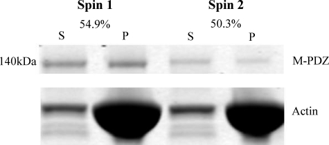
m ATP (▩), and 60-min incubation (▨). All variations were done at 20 μm actin with results showing no significant effect of any parameter on the myosin saturation curve. C, the effect of ATP on actin binding was further investigated with a range of actin titrations as in A in the presence of 1 mm ATP. Data collected from M-PDZ (●) in the presence of ATP gave a Kd of 1.5 ± 0.1 μm and saturation at 48.2 ± 14.5%. Data from M-ΔPDZ (○) resulted in a Kd of 1.0 ± 0.1 μm and saturation at 81.3 ± 8.9%. Conditions were as described in A except for the inclusion of 1 mm ATP.
FIGURE 7.
FIGURE 7.
Evidence for two conformational states of myosin-18 motor. Myosin-18-MMD proteins were sedimented with 20 μm actin as in Fig. 6. The left two lanes show the supernatant (S) and pellet (P) fractions from a representative experiment using M-PDZ. The supernatant from this experiment was mixed with 20 μm actin, and a second sedimentation was performed. The supernatant and pellet from this experiment are shown in the right two lanes. The fraction of actin bound in the first sedimentation was 54.9%, and the fraction bound in the second sedimentation was 50.3%. Ionic conditions were as described in Fig. 6.
FIGURE 8.
FIGURE 8.
Attenuation of skeletal muscle myosin-2 heavy meromyosin in vitro motility by Drosophila myosin-18. Varying ratios of rabbit skeletal muscle myosin-2 heavy meromyosin to M-PDZ (●) or M-ΔPDZ (○) were mixed together with the total myosin concentration held constant at 0.2 mg/ml. Motility was assayed at 30 °C in buffer containing final concentrations of 50 mm KCl, 20 mm MOPS (pH 7.4), 5 mm MgCl2, 0.1 mm EGTA, 1 mm ATP, 25 μg/ml glucose oxidase, 45 μg/ml catalase, 2.5 mg/ml glucose, and 50 mm DTT. Centroid tracking of at least 15 filaments was performed and analyzed with the CellTrak program. Noise within the motility setup was determined to be 0.066 ± 0.035 μm/s (n = 26) by imaging immobile actin filaments bound to a surface coated with 0.2 mg/ml SkHMM in the absence of ATP.
FIGURE 9.
FIGURE 9.
Optical trapping analysis of single molecule interactions. A and B, three-bead assays with oscillations show brief interactions between M-PDZ (A) or M-ΔPDZ (B) and rhodamine-phalloidin stabilized F-actin. Arrows point to attachment events. C and D, lifetime data of myosin-18 motor interactions longer than 10 ms collected in the optical trap were fitted to a single exponential curve. M-PDZ (C) yielded a detachment rate of 94.2 ± 1.0 s−1, and M-ΔPDZ (D) yielded a detachment rate of 57.0 ± 0.6 s−1. E and F, fitting the displacement data collected from the optical trap during each actomyosin-18 interaction to a Gaussian distribution yielded histograms centered at −0.98 ± 1.6 nm for M-PDZ (E) and at −0.03 ± 1.35 nm for M-ΔPDZ (F). Data were collected at 22 °C in a buffer containing 25 mm KCl, 25 mm imidazole (pH 7.4), 4 mm MgCl2, 1 mm EGTA, 2 mm creatine phosphate, 50 mm DTT, 10 μm ATP, 0.1 mg/ml creatine phosphokinase, 3 mg/ml glucose, 0.1 mg/ml glucose oxidase, and 0.02 mg/ml catalase.
FIGURE 10.
FIGURE 10.
Unique amino acid sequence features of Drosophila myosin-18. A, sequence alignments of amino acid residues from the P-loop, Switch I (upper alignment), and Switch II (middle alignment) regions of Drosophila myosin-18 in comparison with consensus sequences from these regions and sequences from motor domains of Dd Myo2 and Mus musculus myosin-18A (Mm myo18A). The lower alignment demonstrates the presence of a 21-residue insertion for D. melanogaster myosin-18 (Dm Myo18) and a 29-residue insertion for Mus musculus myosin-18A between the region bounded by amino acids Gln662 and Leu663 in Dd Myo2. Alignments also reveal amino acid insertions in multiple locations, including a 6-residue insertion between Dd Myo2 residues Asn464 and Ser465. Bold residues are discussed in the text. B and C, using ribbon diagrams of the crystal structure of the motor domain of Dd Myo2 (Protein Data Bank code 1MMD; ADP, blue), the two D. melanogaster myosin-18 amino acid extensions in A are illustrated by highlighting the flanking Dd Myo2 amino acids. The 6-residue insertion between Dd Myo2 Asn464 and Ser465 (red) is located at the end of Switch II, whereas the 21-residue insertion between Gln662 and Leu663 (yellow) is located before the SH2 helix of the motor. D, space-filling model of the crystal structure of the same region as in C. The Gln662-Leu663 insertion can be seen on the surface of the motor, suggesting it to be a large surface loop that may potentially change how rearrangements of the motor domain occur during the kinetic cycle. The Asn464-Ser465 insertion, however, cannot be seen clearly in the space-filling model, suggesting that the 6-residue insertion would be buried within the motor structure and could potentially change the dynamics of cleft closure and perhaps produce a well defined motor structure with low nucleotide affinity and weakened affinity for actin.

References

    1. Odronitz F., Kollmar M. (2007) Genome Biol. 8, R196 - PMC - PubMed
    1. Furusawa T., Ikawa S., Yanai N., Obinata M. (2000) Biochem. Biophys. Res. Commun. 270, 67–75 - PubMed
    1. Doyle D. A., Lee A., Lewis J., Kim E., Sheng M., MacKinnon R. (1996) Cell 85, 1067–1076 - PubMed
    1. Nishioka M., Kohno T., Tani M., Yanaihara N., Tomizawa Y., Otsuka A., Sasaki S., Kobayashi K., Niki T., Maeshima A., Sekido Y., Minna J. D., Sone S., Yokota J. (2002) Proc. Natl. Acad. Sci. U.S.A. 99, 12269–12274 - PMC - PubMed
    1. Ajima R., Kajiya K., Inoue T., Tani M., Shiraishi-Yamaguchi Y., Maeda M., Segawa T., Furuichi T., Sutoh K., Yokota J. (2007) Biochem. Biophys. Res. Commun. 356, 851–856 - PubMed

Publication types

LinkOut - more resources