

RUNX1T1: a novel predictor of liver metastasis in primary pancreatic endocrine neoplasms
- PMID: 21499216
- PMCID: PMC4732279
- DOI: 10.1097/MPA.0b013e3182152bda
RUNX1T1: a novel predictor of liver metastasis in primary pancreatic endocrine neoplasms
Abstract
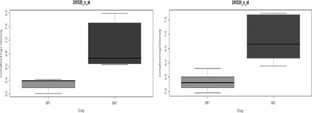
Objectives: Using gene expression profiling on frozen primary pancreatic endocrine tumors (PETs), we discovered RUNX1T1 as a leading candidate progression gene. This study was designed (1) to validate the differential expression of RUNX1T1 protein on independent test sets of metastatic and nonmetastatic PETs and (2) to determine if RUNX1T1 underexpression in primary tumors was predictive of liver metastases.
Methods: Immunohistochemical expression of RUNX1T1 protein was quantified using Allred scores on archival metastatic (n = 13) and nonmetastatic (n = 24) primary adult PET tissues using custom-designed tissue microarrays. Wilcoxon rank sum/Fisher exact tests and receiver operating characteristic curves were used in the data analysis.
Results: Median RUNX1T1 scores were 2 (2-7) and 6 (3-8) in metastatic versus nonmetastatic primaries (P < 0.0001). Eleven of 13 metastatic and 1 of 24 nonmetastatic primaries exhibited RUNX1T1-scores of 4 or less (P < 0.0001). Low RUNX1T1 expression was highly associated with hepatic metastases (P < 0.0001), whereas conventional histological criteria (Ki-67 index, mitotic rate, necrosis) were weakly associated with metastases (P = 0.08-0.15). Considering RUNX1T1 expression (Allred) score of 4 or less to be predictive, the sensitivity to predict hepatic metastases was 85%, with a specificity of 96%.
Conclusions: RUNX1T1 protein is underexpressed in well-differentiated metastatic primary PETs relative to nonmetastatic primaries and emerges as a promising novel biomarker for prediction of liver metastases.
Conflict of interest statement
The authors have no conflict of interest to disclose.
Figures


Similar articles
-
p21 Protein Outperforms Clinico-pathological Criteria in Predicting Liver Metastases in Pancreatic Endocrine Tumors.Cancer Genomics Proteomics. 2023 Nov-Dec;20(6):522-530. doi: 10.21873/cgp.20402. Cancer Genomics Proteomics. 2023. PMID: 37889062 Free PMC article.
-
Met proto-oncogene and insulin-like growth factor binding protein 3 overexpression correlates with metastatic ability in well-differentiated pancreatic endocrine neoplasms.Clin Cancer Res. 2004 Sep 15;10(18 Pt 1):6152-8. doi: 10.1158/1078-0432.CCR-04-0285. Clin Cancer Res. 2004. PMID: 15448002
-
Overexpression of membrane proteins in primary and metastatic gastrointestinal neuroendocrine tumors.Ann Surg Oncol. 2013 Dec;20 Suppl 3(0 3):S739-S746. doi: 10.1245/s10434-013-3318-6. Epub 2013 Oct 10. Ann Surg Oncol. 2013. PMID: 24114056 Free PMC article.
-
EPB41L5 is Associated With the Metastatic Potential of Low-grade Pancreatic Neuroendocrine Tumors.Cancer Genomics Proteomics. 2019 Sep-Oct;16(5):309-318. doi: 10.21873/cgp.20136. Cancer Genomics Proteomics. 2019. PMID: 31467225 Free PMC article.
-
Highlights of the biology of endocrine tumours of the gut and pancreas.Endocr Relat Cancer. 2003 Dec;10(4):427-36. doi: 10.1677/erc.0.0100427. Endocr Relat Cancer. 2003. PMID: 14713255 Review.
Cited by
-
p21 Protein Outperforms Clinico-pathological Criteria in Predicting Liver Metastases in Pancreatic Endocrine Tumors.Cancer Genomics Proteomics. 2023 Nov-Dec;20(6):522-530. doi: 10.21873/cgp.20402. Cancer Genomics Proteomics. 2023. PMID: 37889062 Free PMC article.
-
Detection of early stage pancreatic cancer using 5-hydroxymethylcytosine signatures in circulating cell free DNA.Nat Commun. 2020 Oct 19;11(1):5270. doi: 10.1038/s41467-020-18965-w. Nat Commun. 2020. PMID: 33077732 Free PMC article.
-
C/EBPβ regulates homeostatic and oncogenic gastric cell proliferation.J Mol Med (Berl). 2016 Dec;94(12):1385-1395. doi: 10.1007/s00109-016-1447-7. Epub 2016 Aug 13. J Mol Med (Berl). 2016. PMID: 27522676 Free PMC article.
-
Differentially expressed microRNAs in lung adenocarcinoma invert effects of copy number aberrations of prognostic genes.Oncotarget. 2018 Jan 8;9(10):9137-9155. doi: 10.18632/oncotarget.24070. eCollection 2018 Feb 6. Oncotarget. 2018. PMID: 29507679 Free PMC article.
-
Nuclear imaging of neuroendocrine tumors with unknown primary: why, when and how?Eur J Nucl Med Mol Imaging. 2015 Jun;42(7):1144-55. doi: 10.1007/s00259-015-3027-4. Epub 2015 Mar 13. Eur J Nucl Med Mol Imaging. 2015. PMID: 25771906 Review.
References
-
- Kulke M. Advances in the treatment of neuroendocrine tumors. Curr Treat Options Oncol. 2005;6:397–409. - PubMed
-
- Oberg K, Eriksson B. Medical treatment of neuroendocrine gut and pancreatic tumors. Acta Oncol. 1989;28:425–431. - PubMed
-
- Modlin IM, Sandor A. An analysis of 8305 cases of carcinoid tumors. Cancer. 1997;79:813–829. - PubMed
-
- DeLellis R, Lloyd R, Heitz P, et al. Pathology and Genetics of Tumors of Endocrine Organs. Lyon, France: IARC Press; 2004.
-
- Hochwald SN, Zee S, Conlon KC, et al. Prognostic factors in pancreatic endocrine neoplasms: an analysis of 136 cases with a proposal for low-grade and intermediate-grade groups. J Clin Oncol. 2002;20:2633–2642. - PubMed
Publication types
MeSH terms
Substances
Grants and funding
LinkOut - more resources
Full Text Sources
Medical
