

Proteasome inhibitor MG-132 induces C6 glioma cell apoptosis via oxidative stress
- PMID: 21499287
- PMCID: PMC4002511
- DOI: 10.1038/aps.2011.16
Proteasome inhibitor MG-132 induces C6 glioma cell apoptosis via oxidative stress
Abstract
Aim: Proteasome inhibitors have been found to suppress glioma cell proliferation and induce apoptosis, but the mechanisms are not fully elucidated. In this study we investigated the mechanisms underlying the apoptosis induced by the proteasome inhibitor MG-132 in glioma cells.
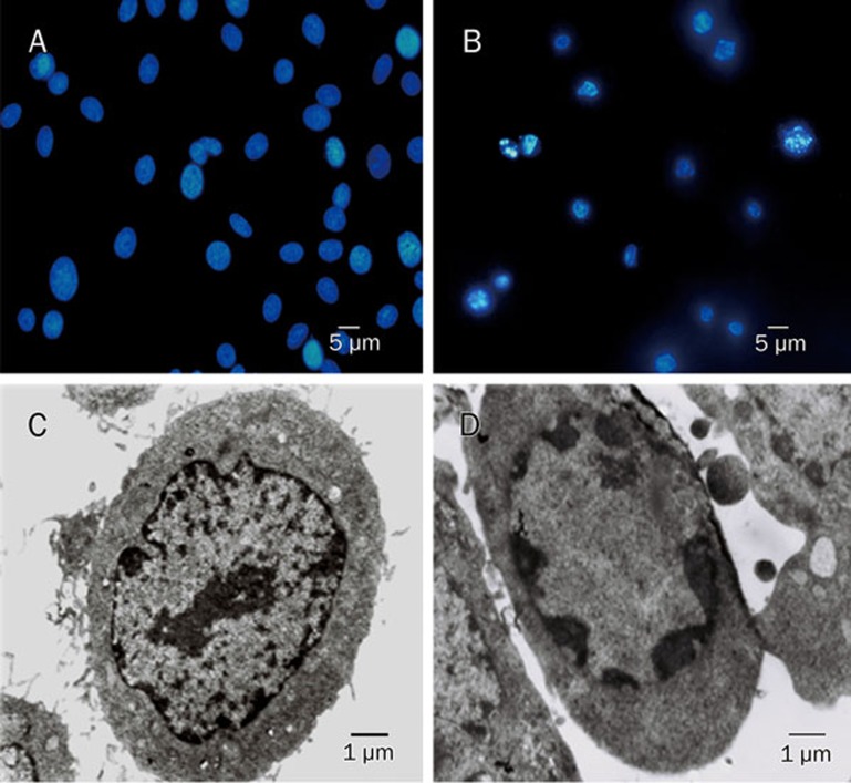
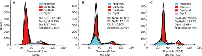
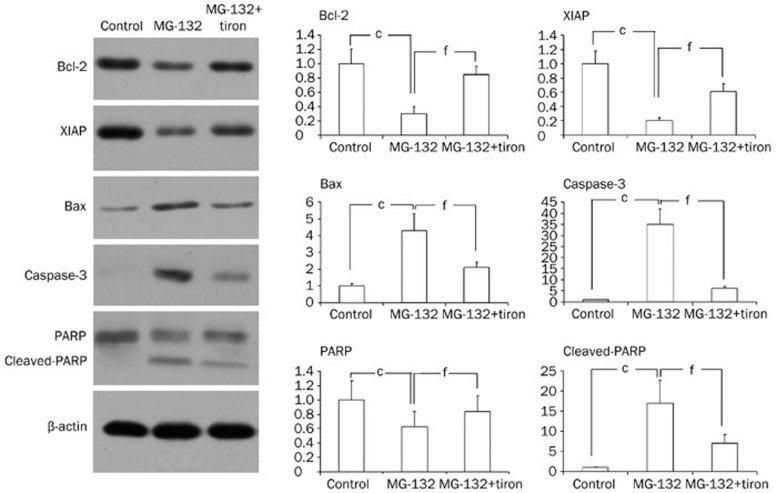
Methods: C6 glioma cells were used. MTT assay was used to analyze cell proliferation. Proteasome activity was assayed using Succinyl-LLVY-AMC, and intracellular ROS level was evaluated with the redox-sensitive dye DCFH-DA. Apoptosis was detected using fluorescence and transmission electron microscopy as well as flow cytometry. The expression of apoptosis-related proteins was investigated using Western blot analysis.
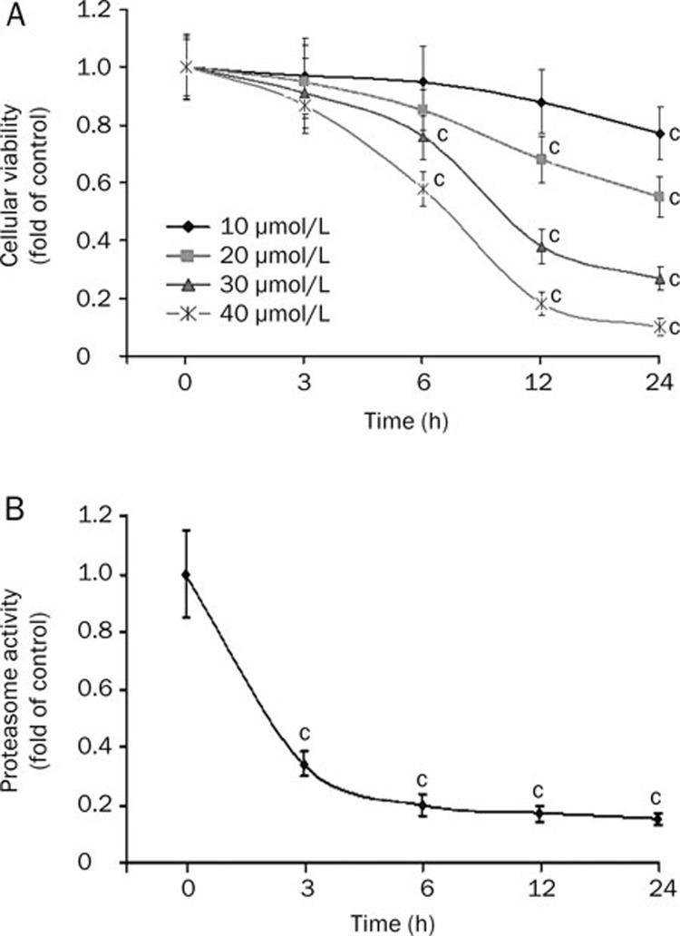
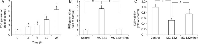
Results: MG-132 inhibited C6 glioma cell proliferation in a time- and dose-dependent manner (the IC(50) value at 24 h was 18.5 μmol/L). MG-132 (18.5 μmol/L) suppressed the proteasome activity by about 70% at 3 h. It induced apoptosis via down-regulation of antiapoptotic proteins Bcl-2 and XIAP, up-regulation of pro-apoptotic protein Bax and caspase-3, and production of cleaved C-terminal 85 kDa PARP). It also caused a more than 5-fold increase of reactive oxygen species. Tiron (1 mmol/L) effectively blocked oxidative stress induced by MG-132 (18.5 μmol/L), attenuated proliferation inhibition and apoptosis in C6 glioma cells, and reversed the expression pattern of apoptosis-related proteins.
Conclusion: MG-132 induced apoptosis of C6 glioma cells via the oxidative stress.
Figures
References
-
- Rubinsztein DC. The roles of intracellular protein-degradation pathways in neurodegeneration. Nature. 2006;443:780–6. - PubMed
-
- Hershko A. The ubiquitin system for protein degradation and some of its roles in the control of the cell division cycle. Cell Death Differ. 2005;12:1191–7. - PubMed
-
- Sorolla A, Yeramian A, Dolcet X, de Santos AMPérez, Llobet D, Schoenenberger JA, et al. Effect of proteasome inhibitors on proliferation and apoptosis of human cutaneous melanoma-derived cell lines. Br J Dermatol. 2008;158:496–504. - PubMed
-
- Momose I, Iijima M, Kawada M, Ikeda D. A new proteasome inhibitor, TP-110, induces apoptosis in human prostate cancer PC-3 cells. Biosci Biotechnol Biochem. 2007;71:1036–43. - PubMed
-
- Yin D, Zhou H, Kumagai T, Liu G, Ong JM, Black KL, et al. Proteasome inhibitor PS-341 causes cell growth arrest and apoptosis in human glioblastoma multiforme (GBM) Oncogene. 2005;24:344–54. - PubMed
Publication types
MeSH terms
Substances
LinkOut - more resources
Full Text Sources
Research Materials
