. 2011 Oct 6;30(40):4175-84.
doi: 10.1038/onc.2011.126. Epub 2011 Apr 18.

Molecular targeting of CSN5 in human hepatocellular carcinoma: a mechanism of therapeutic response

Affiliations

Molecular targeting of CSN5 in human hepatocellular carcinoma: a mechanism of therapeutic response

Y-H Lee et al. Oncogene. .

Abstract

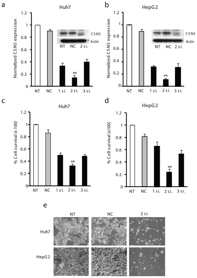
Development of targeted therapy for hepatocellular carcinoma (HCC) remains a major challenge. We have recently identified an elevated expression of the fifth subunit of COP9 signalosome (CSN5) in early HCC as compared with dysplastic stage. In the present study, we explored the possibility of CSN5 being a potential therapeutic target for HCC. Our results show that CSN5 knockdown by small-interfering (si) RNA caused a strong induction of apoptosis and inhibition of cell-cycle progression in HCC cells in vitro. The down-regulation of CSN5 was sufficient to interfere with CSN function as evidenced by the accumulation of neddylated Cullin 1 and changes in the protein levels of CSN-controlled substrates SKP2, p53, p27 and nuclear factor-κB, albeit to a different degree depending on the HCC cell line, which could account for the CSN5 knockdown phenotype. The transcriptomic analysis of CSN5 knockdown signature showed that the anti-proliferative effect was driven by a common subset of molecular alterations including down-regulation of cyclin-dependent kinase 6 (CDK6) and integrin β1 (ITGB1), which were functionally interconnected with key oncogenic regulators MYC and TGFβ1 involved in the control of proliferation, apoptotic cell death and HCC progression. Consistent with microarray analysis, western blotting revealed that CSN5 depletion increased phosphorylation of Smad 2/3, key mediators of TGFβ1 signaling, decreased the protein levels of ITGB1, CDK6 and cyclin D1 and caused reduced expression of anti-apoptotic Bcl-2, while elevating the levels of pro-apoptotic Bak. A chemically modified variant of CSN5 siRNA was then selected for in vivo application based on the growth inhibitory effect and minimal induction of unwanted immune response. Systemic delivery of the CSN5 3/8 variant by stable-nucleic-acid-lipid particles significantly suppressed the tumor growth in Huh7-luc+ orthotopic xenograft model. Taken together, these results indicate that CSN5 has a pivotal role in HCC pathogenesis and maybe an attractive molecular target for systemic HCC therapy.


Conflict of interest statement

Conflict of Interest

The authors declare no conflict of interest.

Figures

Figure 1
Figure 1
siRNA knockdown of CSN5 inhibits growth of HCC cells. (ab) Downregulation of CSN5 mRNA and protein (insets) in Huh7 (a) or HepG2 (b) cells treated with CSN5-specific siRNA. Total RNAs and whole cell lysates were extracted at 48 h after treatment with 15 nM of siRNAs. CSN5 mRNA expression levels were calculated relative to GAPDH and normalized to untreated control. Actin protein was used as a loading control. The data are shown as means ± S.D. of triplicate experiments (**P < 0.01; Bootstrap Test). (ce) Growth inhibition of Huh7 (c) or HepG2 (d) cells as measured by an MTT assay or microscopic observation (e) (X100 original magnification) 4 d after transfection. The cell survival was calculated relative to untreated cells. The data are shown as means ± S.D. of triplicate experiments (**P < 0.01, Bootstrap t-test). NT, no treatment; NC, negative control siRNA; 1si., CSN5-1siRNA; 2si., CSN5-2siRNA; 3si., CSN5-3siRNA.
Figure 2
Figure 2
Biological effects of CSN5 siRNA knockdown in HCC cell lines. (a) Cell cycle analysis of HCC cells treated with CSN5-2siRNA for 2 d. (b) Detection of apoptosis 3 d after transfection with CSN5-2siRNA. (c) Effect of CSN5-2siRNA on the proliferation in various HCC cells. The cell survival was calculated relative to NCsiRNA treatment. All statistical analysis was performed using Bootstrap t-test. The data shown are means ± S.D. of triplicate experiments. **, P < 0.01.
Figure 3
Figure 3
Transcriptomic analysis in gene expression following CSN5 knockdown. Huh7 and HepG2 cells were transfected with 15 nM of CSN5-2siRNA and analyzed for changes in gene expression at 48 h following treatment by illumina microarray. The means of the intensity log ratios in CSN5 siRNA-treated cells were calculated relative to the NCsiRNA-treated cells. (a) Fold-changes in expressions of CSN isoforms. (b) Heat-map overview of 127 genes commonly dysregulated in both Huh7 and HepG2. The scale bar (log2-transformed scale) indicate high (red) and low (green) expression levels. **, P <0.05 by Bootstrap t-test. (c–d) Expression targets of MYC (c) or TGFβ1 (d) in CSN5 knockdown signature. Up- and downregulated genes are indicated in red and green, respectively. Genes in gray are associated with the regulated genes.
Figure 4
Figure 4
Molecular mechanisms of therapeutic response to CSN5 targeting. (a) Western blot analysis of CSN components, neddylated cullins and CRL substrates in Huh7 and HepG2 cells treated with the indicated siRNA for 48 h. (b) Western blot analysis of phosphorylated Smad2/3 and the indicated proteins functionally involved in the cell proliferation or apoptosis. Actin was included as a loading control. (c) Growth inhibition of Huh7 or HepG2 cells as measured by an MTT assay 4 d after transfection of PDGFβ siRNA. (d,e) Induction of apoptosis (d) and caspase-3 (e) after transfection with PDGFβ siRNA. All data were calculated relative to NCsiRNA-treated cells. The data are shown as means ± S.D. of triplicate experiments (**P < 0.01, *P < 0.05, Bootstrap t-test). Pβsi., PDGFβ siRNA
Figure 5
Figure 5
Selection of CSN5 3/8 for in vivo application based on the inhibition of tumor cell growth and minimal cytokine induction. (a) Inhibition of Huh7-luc+ cell growth after transfection with 15 nM of SNALP-formulated CSN5-2 (native) or its modified variants (CSN5-3/6~9, CSN5-4/6~9, CSN5-5/6~9). The siRNA transfectants were examined by MTT assay at 4 d after treatment. **, P < 0.01 (n=3) by Bootstrap t-test. SNALP-Luc, SNALP-fromulated siRNA targeting luciferase. (b) Quantification of IL-6 level after CSN5 targeting. Culture supernatants of Flt3L-derived dendrocytes were assayed for IL-6 using ELISA at 24 h after siRNA treatment. The data are shown as the means ± S.D. of triplicate experiments.
Figure 6
Figure 6
Systemic delivery of CSN5 3/8siRNA by SNALP suppresses human HCC growth in a mouse model of orthotopic transplantation. SCID/beige mice received 0.5×10⁶ Huh7-luc+ cells through intrasplenic injection resulting in tumorous growth in the liver. Mice were randomly assigned either to control (SNALP-βgal478) or treatment (SNALP-CSN5 3/8) group based on the intensity of bioluminescence before initiation of siRNA therapy. Two mg/kg of SNALP-βgal478 and SNALP-CSN5 3/8 were injected into tail vein at 8, 11, 14 and 18 days. (a) Representative in vivo bioluminescence images of Huh7-xenografts. Images were set at the same pseudocolor scale to show the relative bioluminescence changes over time. (b) Quantification of bioluminescence. The total flux is plotted as photon/second. **, P < 0.01, (n=8 vs. n=6) by Mann-Whitney U-test. (c) Histopathological evaluation. Shown are representative photos of gross liver morphology and histopathology at 28 days after transplantation. H&E staining, original magnification X50. (d) Liver-to-body weight ratios. The data are shown as the means ± S.D. in each treatment group. **P < 0.01, Mann-Whitney U-test.

References

    1. Adler AS, Lin M, Horlings H, Nuyten DS, van de Vijver MJ, Chang HY. Genetic regulators of large-scale transcriptional signatures in cancer. Nat Genet. 2006;38:421–430. - PMC - PubMed
    1. Adler AS, Littlepage LE, Lin M, Kawahara TL, Wong DJ, Werb Z, et al. CSN5 isopeptidase activity links COP9 signalosome activation to breast cancer progression. Cancer Res. 2008;68:506–515. - PMC - PubMed
    1. Arsura M, Cavin LG. Nuclear factor-kappaB and liver carcinogenesis. Cancer Lett. 2005;229:157–169. - PubMed
    1. Avila MA, Berasain C, Torres L, Martín-Duce A, Corrales FJ, Yang H, et al. Reduced mRNA abundance of the main enzymes involved in methionine metabolism in human liver cirrhosis and hepatocellular carcinoma. J Hepatol. 2000;33:907–914. - PubMed
    1. Bae MK, Ahn MY, Jeong JW, Bae MH, Lee YM, Bae SK, et al. Jab1 interacts directly with HIF-1alpha and regulates its stability. J Biol Chem. 2002;277:9–12. - PubMed

Publication types

MeSH terms

Substances