

Hypoxia-induced Jagged2 promotes breast cancer metastasis and self-renewal of cancer stem-like cells
- PMID: 21499308
- PMCID: PMC3145824
- DOI: 10.1038/onc.2011.122
Hypoxia-induced Jagged2 promotes breast cancer metastasis and self-renewal of cancer stem-like cells
Abstract
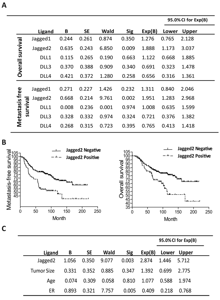
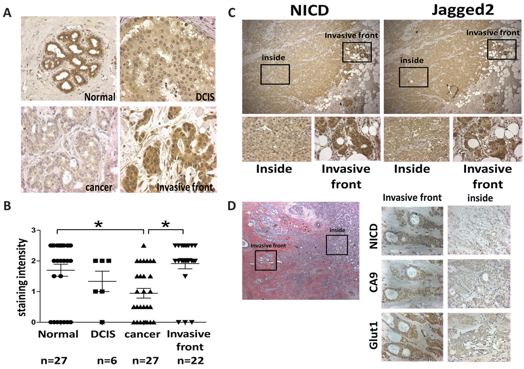
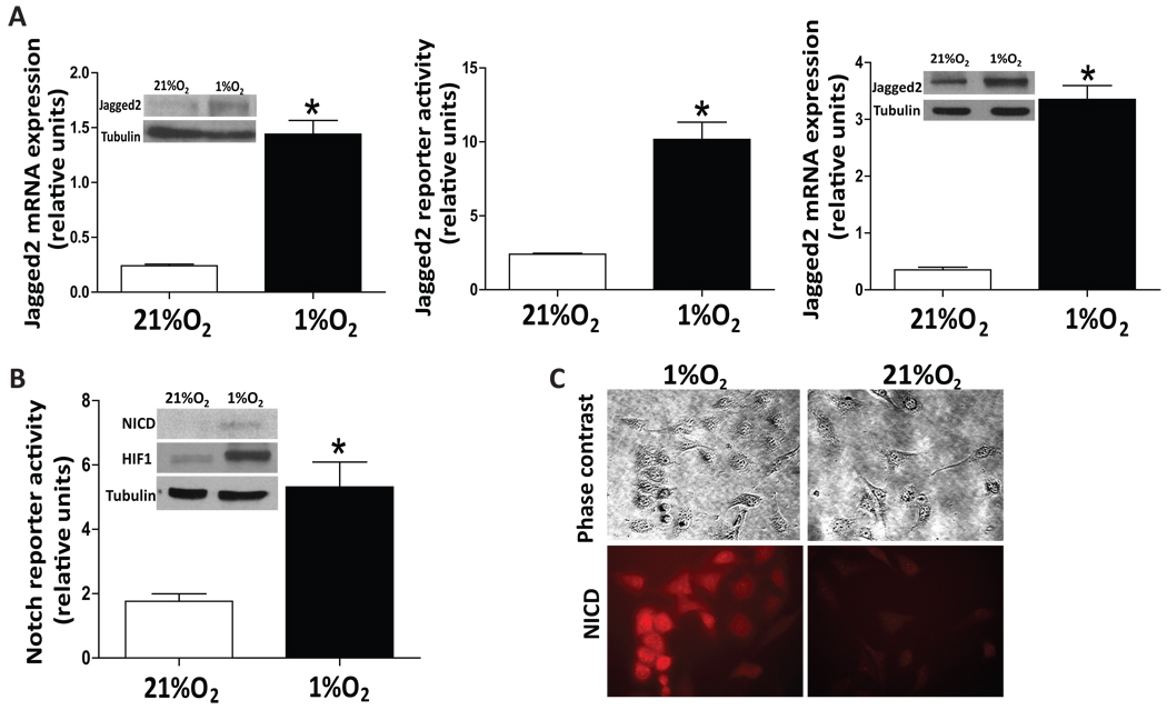
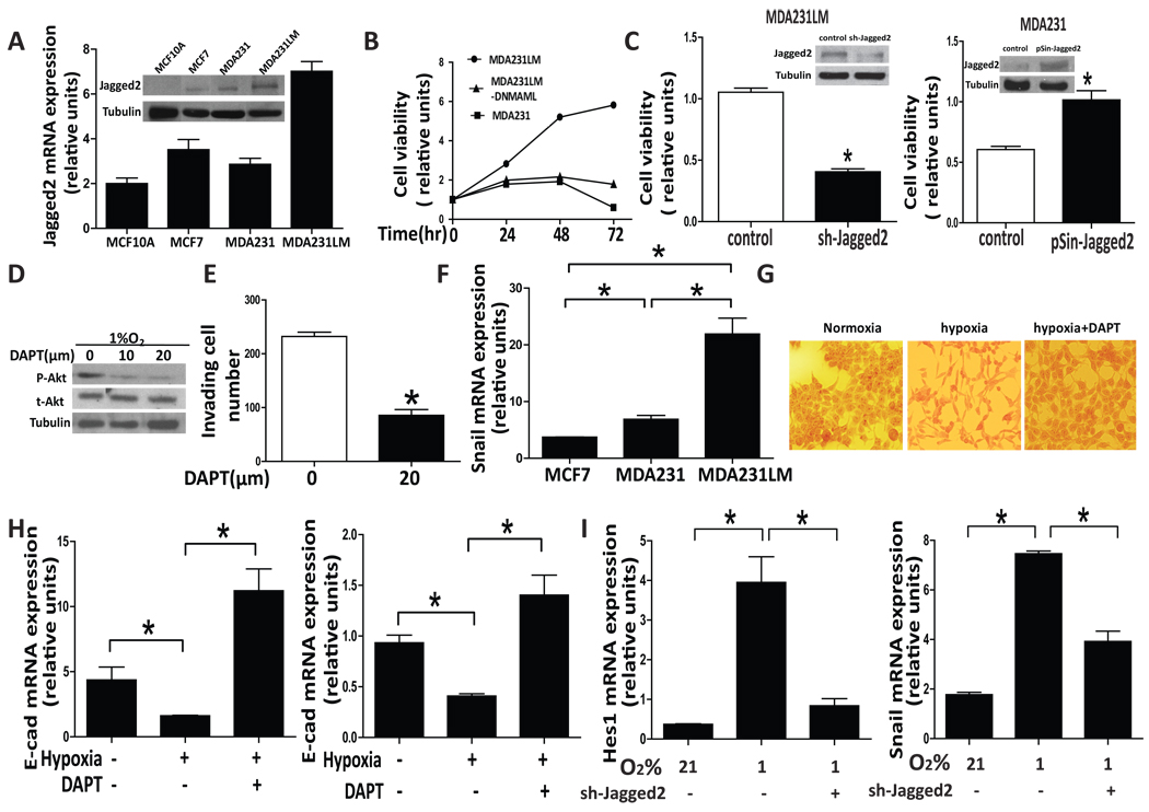
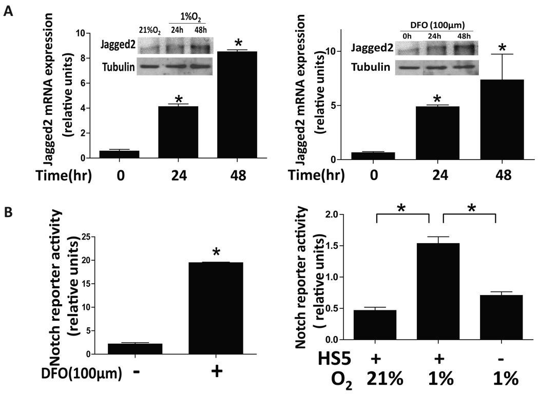
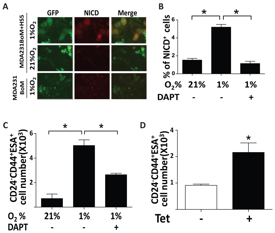
Notch signaling is often and aberrantly activated by hypoxia during tumor progression; however, the exact pathological role of hypoxia-induced Notch signaling in tumor metastasis is as yet poorly understood. In this study, we aimed to define the mechanism of Notch-ligand activation by hypoxia in both primary tumor and bone stromal cells in the metastatic niche and to clarify their roles in tumor progression. We have analyzed the expression profiles of various Notch ligands in 779 breast cancer patients in GEO database and found that the expression of Jagged2 among all five ligands is most significantly correlated with the overall- and metastasis-free survival of breast cancer patients. The results of our immunohistochemical (IHC) analysis for Jagged2 in 61 clinical samples also revealed that both Jagged2 and Notch signaling were strongly upregulated at the hypoxic invasive front. Activation of Jagged2 by hypoxia in tumor cells induced EMT and also promoted cell survival in vitro. Notably, a γ-secretase inhibitor significantly blocked Notch-mediated invasion and survival under hypoxia by promoting expression of E-cadherin and inhibiting Akt phosphorylation. Importantly, Jagged2 was also found to be upregulated in bone marrow stroma under hypoxia and promoted the growth of cancer stem-like cells by activating their Notch signaling. Therefore, hypoxia-induced Jagged2 activation in both tumor invasive front and normal bone stroma has a critical role in tumor progression and metastasis, and Jagged2 is considered to be a valuable prognostic marker and may serve as a novel therapeutic target for metastatic breast cancer.
Conflict of interest statement
The authors declare no conflict of interest.
Figures
Similar articles
-
The NOTCH ligand JAGGED2 promotes pancreatic cancer metastasis independent of NOTCH signaling activation.Mol Cancer Ther. 2015 Jan;14(1):289-97. doi: 10.1158/1535-7163.MCT-14-0501. Epub 2014 Oct 28. Mol Cancer Ther. 2015. PMID: 25351917
-
Reactive astrocytes promote the metastatic growth of breast cancer stem-like cells by activating Notch signalling in brain.EMBO Mol Med. 2013 Mar;5(3):384-96. doi: 10.1002/emmm.201201623. EMBO Mol Med. 2013. PMID: 23495140 Free PMC article.
-
Estrogen-dependent DLL1-mediated Notch signaling promotes luminal breast cancer.Oncogene. 2019 Mar;38(12):2092-2107. doi: 10.1038/s41388-018-0562-z. Epub 2018 Nov 15. Oncogene. 2019. PMID: 30442981 Free PMC article.
-
Notch Signaling in Breast Cancer: A Role in Drug Resistance.Cells. 2020 Sep 29;9(10):2204. doi: 10.3390/cells9102204. Cells. 2020. PMID: 33003540 Free PMC article. Review.
-
Blockade of Jagged/Notch pathway abrogates transforming growth factor β2-induced epithelial-mesenchymal transition in human retinal pigment epithelium cells.Curr Mol Med. 2014 May;14(4):523-34. doi: 10.2174/1566524014666140331230411. Curr Mol Med. 2014. PMID: 24694299 Review.
Cited by
-
Regulation of epithelial-mesenchymal transition by tumor microenvironmental signals and its implication in cancer therapeutics.Semin Cancer Biol. 2023 Jan;88:46-66. doi: 10.1016/j.semcancer.2022.12.002. Epub 2022 Dec 13. Semin Cancer Biol. 2023. PMID: 36521737 Free PMC article. Review.
-
Pannexin1 Is Associated with Enhanced Epithelial-To-Mesenchymal Transition in Human Patient Breast Cancer Tissues and in Breast Cancer Cell Lines.Cancers (Basel). 2019 Dec 7;11(12):1967. doi: 10.3390/cancers11121967. Cancers (Basel). 2019. PMID: 31817827 Free PMC article.
-
Targeting Epigenetic Modifiers of Tumor Plasticity and Cancer Stem Cell Behavior.Cells. 2022 Apr 21;11(9):1403. doi: 10.3390/cells11091403. Cells. 2022. PMID: 35563709 Free PMC article. Review.
-
Systematic expression analysis of ligand-receptor pairs reveals important cell-to-cell interactions inside glioma.Cell Commun Signal. 2019 May 22;17(1):48. doi: 10.1186/s12964-019-0363-1. Cell Commun Signal. 2019. PMID: 31118022 Free PMC article.
-
Targeting HIF-1α by Natural and Synthetic Compounds: A Promising Approach for Anti-Cancer Therapeutics Development.Molecules. 2022 Aug 15;27(16):5192. doi: 10.3390/molecules27165192. Molecules. 2022. PMID: 36014432 Free PMC article. Review.
References
-
- Artavanis-Tsakonas S, Rand MD, Lake RJ. Science. 1999;284:770–776. - PubMed
-
- Bandyopadhyay S, Wang Y, Zhan R, Pai SK, Watabe M, Iiizumi M, Furuta E, Mohinta S, Liu W, Hirota S, Hosobe S, Tsukada T, Miura K, Takano Y, Saito K, Commes T, Piquemal D, Hai T, Watabe K. Cancer Res. 2006;66:11983–11990. - PubMed
-
- Cejudo-Martin P, Johnson RS. Dev Cell. 2005;9:575–576. - PubMed
Publication types
MeSH terms
Substances
Grants and funding
LinkOut - more resources
Full Text Sources
Other Literature Sources
Medical
