. 2011 May;10(5):389-97.
doi: 10.1038/nmat2992. Epub 2011 Apr 17.

The targeted delivery of multicomponent cargos to cancer cells by nanoporous particle-supported lipid bilayers

Affiliations

The targeted delivery of multicomponent cargos to cancer cells by nanoporous particle-supported lipid bilayers

Carlee E Ashley et al. Nat Mater. 2011 May.

Erratum in

  • Nat Mater. 2011 Jun;10(6):476

Abstract

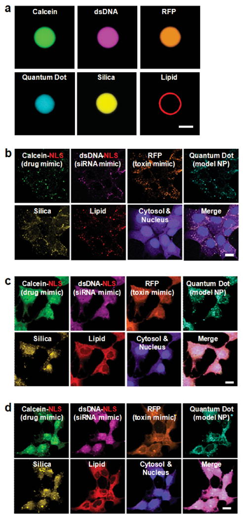
Encapsulation of drugs within nanocarriers that selectively target malignant cells promises to mitigate side effects of conventional chemotherapy and to enable delivery of the unique drug combinations needed for personalized medicine. To realize this potential, however, targeted nanocarriers must simultaneously overcome multiple challenges, including specificity, stability and a high capacity for disparate cargos. Here we report porous nanoparticle-supported lipid bilayers (protocells) that synergistically combine properties of liposomes and nanoporous particles. Protocells modified with a targeting peptide that binds to human hepatocellular carcinoma exhibit a 10,000-fold greater affinity for human hepatocellular carcinoma than for hepatocytes, endothelial cells or immune cells. Furthermore, protocells can be loaded with combinations of therapeutic (drugs, small interfering RNA and toxins) and diagnostic (quantum dots) agents and modified to promote endosomal escape and nuclear accumulation of selected cargos. The enormous capacity of the high-surface-area nanoporous core combined with the enhanced targeting efficacy enabled by the fluid supported lipid bilayer enable a single protocell loaded with a drug cocktail to kill a drug-resistant human hepatocellular carcinoma cell, representing a 10(6)-fold improvement over comparable liposomes.


Figures

Figure 1
Figure 1. Schematic illustration of the nanoporous particle-supported lipid bilayer, depicting the disparate types of therapeutic and diagnostic agents that can be loaded within the nanoporous silica core, as well as the ligands that can be displayed on the surface of the SLB
Targeting and fusogenic peptides are chemically conjugated to phosphatidylethanolamine (DOPE or DPPE), present in the SLB at 1-5 wt%, via a heterobifunctional crosslinker with a polyethylene glycol (PEG) spacer arm (n = 24). The SLB, composed of either fluid (DOPC) or non-fluid (DPPC) zwitterionic lipids with 30 wt% cholesterol, is further modified with 5 wt% PEG-2000 PE to enhance colloidal stability and decrease non-specific interactions.
Figure 2
Figure 2. Physical and biophysical characteristics of protocells
(a) Cryogenic TEM image of the protocell, showing the nanoporous core and the SLB (∼4-nm thick). Particle sizes reflect those naturally generated by the aerosol-assisted self-assembly process; particles were separated into a narrow distribution centered around ∼100-nm for all surface binding, internalization, and delivery experiments (see Supplementary Fig. 1). Scale bar = 25 nm. (b) Temperature-dependent FRAP of NBD-labeled DPPC bilayers (green) supported on nanoporous (○) or solid (●) spherical silica particles. Inset: normalized fluorescence recovery in the photobleached region (blue circle) was determined by dividing the fluorescence intensity (FI) in region of interest 1 (ROI1) by the FI in ROI2 to account for photobleaching that occurred during the recovery period. Scale bar = 5 μm.
Figure 3
Figure 3. Schematic depicting the successive steps of multivalent binding and internalization of targeted protocells, followed by endosomal escape and nuclear localization of protocell-encapsulated cargo
DOPC protocells [1] bind to HCC with high affinity due to recruitment of SP94 targeting peptides (magenta) to the cell surface, [2] become internalized via receptor-mediated endocytosis, and [3] release their cargo into the cytosol upon endosome acidification and protonation of the H5WYG fusogenic peptide (blue). Cargos modified with a NLS are transported through the nuclear pore complex and become concentrated in the nucleus [4].
Figure 4
Figure 4. Selective binding and internalization characteristics of SP94-targeted protocells
(a) and (b) Dissociation constants (Kd) of SP94-targeted protocells and liposomes for Hep3B (a) and hepatocytes (b) as a function of the average number of SP94 peptides per particle (average SP94 wt% is in parentheses). All surface binding experiments were conducted at 4°C to prevent internalization of targeted protocells and liposomes. All error bars in (a) and (b) represent 95% confidence intervals (1.96 σ) for n = 5. (c) Recruitment of Alexa Fluor® 647-labeled SP94 peptides (white) to the surface of a Hep3B cell when peptides are displayed on a NBD-labeled SLB (green) composed of DOPC (○) or DPPC (●). These data were collected at 4°C to replicate the conditions used to determine Kd values in (a) and (b). Hep3B cells were labeled with CellTracker™ Red CMTPX (red) and Hoechst 33342 (blue). Inset scale bars = 5 μm. (d) and (e) Confocal fluorescence microscopy images of Hep3B (d) and hepatocytes (e) incubated with SP94-targeted protocells for 1 hour at 37°C. Protocells were prepared with Texas Red®-labeled DHPE (red) and Alexa Fluor® 647-labeled nanoporous cores (white); cells were stained with CellTracker™ Green CMFDA (green) and Hoechst 33342 (blue). Cells shown in (d) and (e) are representative of the entire cell population (see Supplementary Table II for population-based internalization data); single cells were selected to enable 3D imaging. Plan (left and center images) and cross-sectional (right image) views of the 3D projection are shown for (d), while the plan view alone is shown for (e). For (d), the merged plan view (left) is shown without the green channel (center) to enable better visualization of lipid (red) and silica (white) moieties. It is important to note that plan views of collapsed projections superimpose all slices in the z-direction, giving the misleading appearance of protocells in the nucleus of (d); this is not the case, however, as is evident in an orthogonal view of the projection (image not shown). All scale bars = 10 μm.
Figure 5
Figure 5. Targeted delivery of multicomponent cargos to the cytosol and nuclei of HCC cells
Alexa Fluor® 532-labeled nanoporous silica cores (yellow) were loaded with a multicomponent mixture of four surrogate cargos: calcein (green), an Alexa Fluor® 647-labeled double-stranded DNA oligonucleotide (magenta), red fluorescent protein (orange), and CdSe/ZnS quantum dots (teal). Cargos were sealed in the cores via fusion of Texas Red®-labeled DOPC liposomes (red) that contained 30 wt% cholesterol and 5 wt% PEG-2000 PE, and the resulting SLBs were modified with 0.015 wt% SP94 and 0.500 wt% H5WYG. Protocells were incubated with Hep3B cells (labeled with CellTracker™ Violet BMQC and Hoechst 33342) for 15 minutes, 4 hours, or 12 hours (respectively) at 37°C to collect the images shown in (b) – (d). (a) Hyperspectral confocal fluorescence microscopy slice (z = ∼5 μm) of a 10-μm protocell, demonstrating uniform loading of the nanoporous silica core and complete encapsulation of the core and cargos within the SLB. Particles 100 times larger than those used for all surface-binding, internalization, and delivery studies were used in this experiment to enable optical imaging and have a 2.5 × 10⁵-fold higher capacity for the multicomponent mixture than protocells (100-150 nm in diameter) used to collect the images shown in (b) – (d). Scale bar = 5 μm. (b) – (d): Hyperspectral confocal fluorescence microscopy was employed to individually track the lipid and silica moieties of DOPC protocells (100-150-nm multimodal core), as well as the four surrogate cargos within the cytosol (purple) and nuclei (blue) of Hep3B cells as a function of time. (b) Within 15 minutes of exposing Hep3B to protocells loaded with the multicomponent mixture, the lipid, silica, and cargo moieties have a punctate appearance, indicating that protocells are localized within endosomes. (c) Within 4 hours, the H5WYG peptide promotes endosomal escape, thereby releasing the lipid, silica, and cargos into the cytosol of the Hep3B cells. (d) Within 12 hours, calcein and the dsDNA oligonucleotide, both of which are modified with a NLS, become concentrated in the nucleus, while the RFP and quantum dots (not modified with a NLS) remain largely localized in the cytosol. Protocells used to collect the images shown in (b) – (d) have a high capacity for the multicomponent mixture: 1010 protocells encapsulate 425 μM of calcein, 7.6 μM of the dsDNA oligonucleotide, 945 nM of RFP, and 1.98 × 10¹³ quantum dots. Scale bars = 20 μm.
Figure 6
Figure 6. Cargo capacity, time-dependent release profiles, and concentration-dependent cytotoxicity of SP94-targeted protocells and liposomes that encapsulate chemotherapeutic drugs
(a) Cargo capacity and cytotoxicity of protocells and liposomes loaded with doxorubicin (DOX). Left axis: the absolute and effective capacities of DOPC protocells, DOPC liposomes, and DSPC liposomes for DOX. Absolute capacity is defined as the concentration of DOX that can be physically encapsulated within 1010 particles, while effective capacity is the concentration of DOX that is released upon endocytosis by Hep3B in a form capable of intercalating nuclear DNA. DOPC protocells, when loaded with a cocktail of DOX, 5-fluorouracil (5-FU), and cisplatin, retain their high absolute and effective capacities. The liposome cocktail is composed of equal volumes of DOX-loaded, 5-FU-loaded, and cisplatin-loaded DSPC liposomes. DSPC liposomes that encapsulate 5-FU have an absolute capacity of 765 nM (per 1010 particles) and were prepared using the reverse-phase evaporation method described by B. Elorza, et al.. DSPC liposomes that encapsulate cisplatin have an absolute capacity of 980 nM (per 1010 particles) and were prepared using the technique described by T. Peleg-Shulman, et al.. Right axis: the number of DOX-loaded protocells or liposomes that must be added to 106 MDR1+ Hep3B cells to kill 90% of the cells in the population (LC90) within 24 hours. (b) The time-dependent release of DOX from DOPC protocells, DSPC liposomes, DOPC liposomes, and nanoporous silica cores when exposed to a simulated body fluid (pH 7.4) at 37°C for 21 days. (c) The time-dependent release of DOX from DOPC protocells, DSPC liposomes, and DOPC liposomes when exposed to a pH 5 citric acid buffer at 37°C for 12 hours. Acidic conditions, which mimic those of the endosome, destabilize the SLB and promote release of DOX from the protocell's nanoporous core. (d) Left axis: the number of MDR1+ Hep3B and hepatocytes that remain viable after exposure to 9.6 μM of free DOX, protocell-encapsulated DOX, or liposomal DOX for 24 hours at 37°C. 9.6 μM is the LC90 value of free DOX when exposed to Hep3B with induced MDR (MDR1+ phenotype) and was, therefore, selected as the standardized drug concentration. Cells were exposed to drugs and drug-loaded nanocarriers for 24 hours since the typical doubling time of HCC is 24-36 hours. Right axis: the number of MDR1+ Hep3B that remain viable after exposure to 2.4 μM of free DOX, protocell-encapsulated DOX, or liposomal DOX for 24 hours at 37°C; 2.4 μM is the LC50 value of free DOX. Sytox® Green nucleic acid stain and Alexa Fluor 647®-labeled annexin V were used to distinguish viable (double-negative) from non-viable (single- or double-positive) cells. All error bars represent 95% confidence intervals (1.96 σ) for n = 3.

Comment in

Similar articles

Cited by

References

    1. Peer D, et al. Nanocarriers as an emerging platform for cancer therapy. Nature Nanotechnology. 2007;2:751–760. - PubMed
    1. Wagner V, Dullaart A, Bock AK, Zweck A. The emerging nanomedicine landscape. Nat Biotech. 2006;24:1211–1217. - PubMed
    1. Nel AE, et al. Understanding biophysicochemical interactions at the nano-bio interface. Nat Mater. 2009;8:543–557. - PubMed
    1. Ferrari M. Cancer nanotechnology: Opportunities and challenges. Nature Reviews Cancer. 2005;5:161–171. - PubMed
    1. Maeda H, Wu J, Sawa T, Matsumura Y, Hori K. Tumor vascular permeability and the EPR effect in macromolecular therapeutics: a review. Journal of Controlled Release. 2000;65:271–284. - PubMed

Publication types