. 2011:6:453-61.
doi: 10.2147/IJN.S17057. Epub 2011 Feb 25.

Electrospun chitosan-graft-poly (ɛ-caprolactone)/poly (ɛ-caprolactone) nanofibrous scaffolds for retinal tissue engineering

Affiliations

Electrospun chitosan-graft-poly (ɛ-caprolactone)/poly (ɛ-caprolactone) nanofibrous scaffolds for retinal tissue engineering

Honglin Chen et al. Int J Nanomedicine. 2011.

Abstract

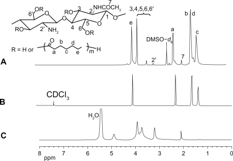
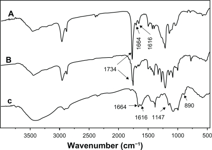
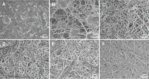
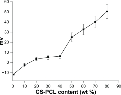
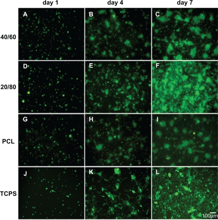
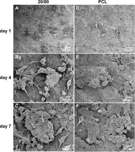
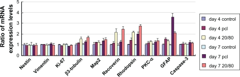
A promising therapy for retinal diseases is to employ biodegradable scaffolds to deliver retinal progenitor cells (RPCs) for repairing damaged or diseased retinal tissue. In the present study, cationic chitosan-graft-poly(ɛ-caprolactone)/polycaprolactone (CS-PCL/PCL) hybrid scaffolds were successfully prepared by electrospinning. Characterization of the obtained nanofibrous scaffolds indicated that zeta-potential, fiber diameter, and the content of amino groups on their surface were closely correlated with the amount of CS-PCL in CS-PCL/PCL scaffolds. To assess the cell-scaffold interaction, mice RPCs (mRPCs) were cultured on the electrospun scaffolds for 7 days. In-vitro proliferation assays revealed that mRPCs proliferated faster on the CS-PCL/PCL (20/80) scaffolds than the other electrospun scaffolds. Scanning electron microscopy and the real-time quantitative polymerase chain reaction results showed that mRPCs grown on CS-PCL/PCL (20/80) scaffolds were more likely to differentiate towards retinal neurons than those on PCL scaffolds. Taken together, these results suggest that CS-PCL/PCL(20/80) scaffolds have potential application in retinal tissue engineering.

Keywords: differentiation; electrospun; proliferation; retinal progenitor cells; tissue engineering.


Figures

Figure 1
Figure 1
Proton nuclear magnetic resonance spectra of CS-PCL (A), PCL (B), and CS (C). Abbreviations: CS, chitosan; CS-PCL, cationic chitosan-graft-poly (ɛ-caprolactone); DMSO, dimethyl sulfoxide; PCL, polycaprolactone.
Figure 2
Figure 2
Fourier-transform infrared spectra of CS-PCL (A), PCL (B), and CS (C). Abbreviations: CS, chitosan; CS-PCL, cationic chitosan-graft-poly (ɛ-caprolactone); PCL, polycaprolactone.
Figure 3
Figure 3
Scanning electron microscopy images of electrospun nanofibous scaffolds prepared from different CS-PCL/PCL weight ratios: CS-PCL (A), CS-PCL/PCL (80/20) (B), CS-PCL/PCL (60/40) (C), CS-PCL/PCL (40/60) (D), CS-PCL/PCL (20/80) (E), and PCL (F). Abbreviations: CS-PCL, cationic chitosan-graft-poly (ɛ-caprolactone); PCL, polycaprolactone.
Figure 4
Figure 4
Zeta-potential curve for electrospun nanofibrous scaffolds (n = 5). Abbreviation: CS-PCL, cationic chitosan-graft-poly (ɛ-caprolactone).
Figure 5
Figure 5
Fluorescent micrograph of mRPC density on CS-PCL/PCL (40/60) (A–C), CS-PCL/PCL (20/80) (D–F), PCL (G–I) nanofibrous scaffolds, and the cell culture plates (TCPS) (J–L) on day 1, 4, and 7 after incubation (green = endogenous GFP expression). Scale bar: 100 μm. Abbreviations: CS-PCL, cationic chitosan-graft-poly (ɛ-caprolactone); GFP, green fluorescent protein; PCL, polycaprolactone; mRPC, mouse retinal progenitor cell; TCPS, tissue culture polystyrene.
Figure 6
Figure 6
Analysis of mRPC proliferation on PCL and CS-PCL/PCL nanofibrous scaffolds and the cell culture plates (TCPS), measured by MTS assay. Notes: Bar represents mean ± standard deviation (n = 4). *P < 0.05. Abbreviations: CS-PCL, cationic chitosan-graft-poly (ɛ-caprolactone); mRPC, mouse retinal progenitor cell; MTS, 3-(4,5-dimethylthiazol-2-yl)-5-(3-carboxymethoxyphenyl)-2-(4-sulfophenyl)-2H-tetrazolium; OD, optical density; PCL, polycaprolactone; TCPS, tissue culture polystyrene.
Figure 7
Figure 7
Scanning electron microscopy images of mRPCs grown on CS-PCL/PCL (20/80) (A–C) and PCL (D–F) nanofibrous scaffolds on day 1 (A, D), 4 (B, E), and 7 (C, F) after incubation. Abbreviations: CS-PCL, cationic chitosan-graft-poly (ɛ-caprolactone); mRPC, mouse retinal progenitor cell; PCL, polycaprolactone.
Figure 8
Figure 8
Gene expression profile of mRPCs grown on CS-PCL/PCL (20/80) and PCL scaffolds on day 4 and 7. Note: The error bars show standard deviation (n = 3). Abbreviations: CS-PCL, cationic chitosan-graft-poly (ɛ-caprolactone); GFAP, glial fibrillary acidic protein; mRPC, mouse retinal progenitor cell; PCL, polycaprolactone; PKC-α, protein kinase C alpha.

References

    1. Margalit E, Sadda SR. Retinal and optic nerve diseases. Artif Organs. 2003;27(11):963–974. - PubMed
    1. Ambati J, Ambati BK, Yoo SH, Ianchulev S, Adamis AP. Age-related macular degeneration: etiology, pathogenesis, and therapeutic strategies. Surv Ophthalmol. 2003;48(3):257–293. - PubMed
    1. Klassen HJ, Ng TF, Kurimoto Y, et al. Multipotent retinal progenitors express developmental markers, differentiate into retinal neurons, and preserve light-mediated behavior. Invest Ophthalmol Vis Sci. 2004;45(11):4167–4173. - PubMed
    1. Redenti S, Neeley WL, Rompani S, et al. Engineering retinal progenitor cell and scrollable poly (glycerol-sebacate) composites for expansion and subretinal transplantation. Biomaterials. 2009;30(20):3405–3414. - PMC - PubMed
    1. Lavik EB, Klassen H, Warfvinge K, Langer R, Young MJ. Fabrication of degradable polymer scaffolds to direct the integration and differentiation of retinal progenitors. Biomaterials. 2005;26(16):3187–3196. - PubMed

Publication types

MeSH terms