

H HRMAS NMR Derived Bio-markers Related to Tumor Grade, Tumor Cell Fraction, and Cell Proliferation in Prostate Tissue Samples
- PMID: 21499438
- PMCID: PMC3076017
- DOI: 10.4137/BMI.S6794
H HRMAS NMR Derived Bio-markers Related to Tumor Grade, Tumor Cell Fraction, and Cell Proliferation in Prostate Tissue Samples
Abstract
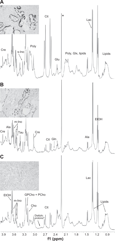
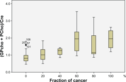
A high-resolution magic angle spinning NMR spectroscopic approach is presented for evaluating the occurrence, amount and aggressiveness of cancer in human prostate tissue samples. Using this technique, key metabolites in malignant and non-malignant samples (n = 149) were identified, and patterns of their relative abundance were analyzed by multivariate statistical methods. Ratios of various metabolites - including (glycerophophorylcholine + phosphorylcholine)/creatine, myo-inositol/scyllo-inositol, scyllo-inositol/creatine, choline/creatine, and citrate/creatine - correlated with: i) for non-malignant tissue samples, the distance to the nearest tumor and its Gleason score and; ii) the fraction of tumor cells present in the sample; and iii) tumor cell proliferation (Ki67 labelling index). This NMR-based approach allows the extraction of information that could be useful for developing novel diagnostic methods for prostate cancer.
Keywords: Gleason score; HRMAS; Ki67; MRSI; inositol; prostate cancer.
Figures
References
-
- Wolf AMD, Wender RC, Etzioni RB, Thompson IM, D’Amico AV, et al. American Cancer Society Guideline for the Early Detection of Prostate Cancer. CA Cancer J Clin. 2010;60:70–98. - PubMed
-
- Chrouser KL, Lieber MM. Extended and saturation needle biopsy for the diagnosis of prostate cancer. Curr Urol Rep. 2004;5(3):226–30. - PubMed
-
- Fleshner N, Klotz L. Role of “saturation biopsy” in the detection of prostate cancer among difficult diagnostic cases. Urology. 2002;60:93–7. - PubMed
-
- Baccala AA, Moussa AS, Elbary AA, et al. Risk factors and predictors of prostate cancer in men with negative repeat saturation biopsy. Uro Today Int J. 2010;3(1)
LinkOut - more resources
Full Text Sources
Other Literature Sources
