. 2011 Apr 1;255(7-8):688-699.
doi: 10.1016/j.ccr.2010.10.016.

Structural, Mechanistic and Coordination Chemistry of Relevance to the Biosynthesis of Iron-Sulfur and Related Iron Cofactors

Affiliations

Structural, Mechanistic and Coordination Chemistry of Relevance to the Biosynthesis of Iron-Sulfur and Related Iron Cofactors

Wenbin Qi et al. Coord Chem Rev. .

Abstract

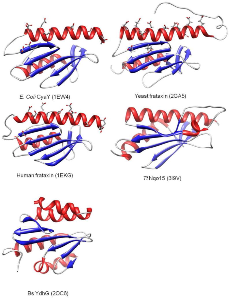
Iron-sulfur clusters are an important class of protein-bound prosthetic center that find wide utility in nature. Roles include electron transfer, enzyme catalysis, protein structure stabilization, and regulation of gene expression as transcriptional and translational sensors. In eukaryotes their biosynthesis requires a complex molecular machinery that is located within the mitochondrion, while bacteria exhibit up to three independent cluster assembly pathways. All of these paths share common themes. This review summarizes some key structural and functional properties of three central proteins dedicated to the Fe-S cluster assembly process: namely, the sulfide donor (cysteine desulfurase); iron donor (frataxin), and the iron-sulfur cluster scaffold protein (IscU/ISU).


Figures

Figure 1
Figure 1
Prototypical structures of the most common iron-sulfur centers. [2Fe-2S], [3Fe-4S] and [4Fe-4S] clusters with cysteines typically providing the additional ligand contacts to iron.
Figure 2
Figure 2
Schematic illustration of the key steps and proteins involved in iron-sulfur cluster biosynthesis. In the first step, IscU/ISU receives iron and sulfide from the iron donor protein frataxin and sulfur donor protein IscS, respectively, and assembles the [2Fe-2S] iron-sulfur cluster. Subsequently the assembled cluster is transferred to an apo target protein.
Figure 3
Figure 3
IscU core structures. Crystallographic and NMR solution studies show very similar core structures for Hi IscU, Mm IscU, Bs IscU, Tt IscU and Aa IscU.
Figure 4
Figure 4
Sequence alignment for IscU-type proteins. Three conserved cysteine residues that are directly coordinated to the cluster are highlighted in yellow. The semi-conserved histidine and corresponding lysine residues are marked as green or purple, respectively. Other possible non-cysteinyl ligands include Asp39 and Ser65 (numbering as in H. influenzae) are marked in blue.
Figure 5
Figure 5
Cluster coordination. Solution and crystal structures of zinc-bound (A and B) or cluster-bound (C) forms of IscU proteins provide evidence that the cluster is ligated by three conserved cysteine residues and a fourth non-cysteinyl ligand. (A) The structure of the zinc-bound Hi IscU shows that the fourth ligand is His106. (B) In the structure of zinc-bound Bs IscU, the fourth ligand is Asp43. (C) Structure of one subunit of the trimeric Aa IscU, which has a bound [2Fe-2S] cluster with His106 (Aa numbering) as the fourth ligand.
Figure 6
Figure 6
Iron transfer from frataxin to ISU. In human ISU, residue His106 appears to play a role in promoting interaction between ISU and the iron donor protein frataxin (schematically illustrated), as well as mediating iron delivery to the cluster binding site [58].
Figure 7
Figure 7
Sequence alignment for frataxin homologs. Conserved acidic residues on the α1-β1 surface that are proposed to be responsible for iron binding are marked as blue.
Figure 8
Figure 8
Structures of E. coli, yeast and human frataxins show an extremely similar core structure. The conserved acidic residues lying on the α1-β1 surface appear to be iron binding sites [99, 100]. Structures of the Tt Nqo15 [107, 108] and Bs YdhG proteins [89] are both observed to be very similar to the frataxin core.
Figure 9
Figure 9
Sulfide transfer from the IscS-IscU complex. (A) Adapted from the crystallographically defined E. coli IscS-IscU complex [47]. IscS is in light green and IscU is in gray. Residues 328-332 of IscS where Cys328 lie are not solved due to the flexibility of this region, shown as red dotted line. The three conserved cysteine residues of IscU where cluster binds are shown in yellow. Cys328 of IscS is at the position that is not directly close to cluster binding site of IscU. (B) Simulated structural model for the complex of H. influenzae IscU monomer with Tm IscS monomer [53]. Tm IscS is depicted in gray, with the blue region comprising a loop, and a portion of an α-helix adjacent to the disordered loop (residues 321-332) that contains the putative active cysteine residue C324 that mediates sulfide delivery. The position of the latter residue is illustrated with the dotted circle, but is not directly observed in the structure as a result of the flexibility of this loop. IscU is represented in yellow, with the red regions depicting the three conserved cysteine residues (C37, C63, and C106), where the asterisk represents C63, which is poised close to the catalytic C324 of Tm IscS.
Figure 10
Figure 10
Sulfide delivery to IscU/ISU. Cleavage of the IscS persulfide bond by reduced NFU, yielding inorganic sulfide, which is positioned adjacent to the iron-loaded IscU cluster binding site, where X is a presumed stabilizing bridging ligand (possibly a conserved carboxylate [48, 57]). Details of how the iron centers are coordinated to the IscU Cys at the time of sulfide delivery remain to be established.
Figure 11
Figure 11
Sequences of Cys-X-X-Cys domains in NFU-type proteins. Hs, Homo sapiens; Mm, Mus musculus; Dm, Drosophila melanogaster; Ce, Caenorhabditis elegans; Av, Azotobacter vinelandii; Sys, Synechocystis PCC6803.

References

    1. Peters JW, Stowell MH, Soltis SM, Finnegan MG, Johnson MK, Rees DC. Biochemistry. 1997;36:1181–1187. - PubMed
    1. Johnson DC, Dean DR, Smith AD, Johnson MK. Annu Rev Biochem. 2005;74:247–281. - PubMed
    1. Volbeda A, Charon MH, Piras C, Hatchikian EC, Frey M, Fontecilla-Camps JC. Nature. 1995;373:580–587. - PubMed
    1. Iwata S, Saynovits M, Link TA, Michel H. Structure. 1996;4:567–579. - PubMed
    1. Robbins AH, Stout CD. Proc Natl Acad Sci U S A. 1989;86:3639–3643. - PMC - PubMed

LinkOut - more resources