. 2011 Mar 1;71(3):261-265.
doi: 10.1016/j.reactfunctpolym.2010.09.008.

Efficient Intracellular Delivery of a Pro-Apoptotic Peptide With A pH-Responsive Carrier

Affiliations

Efficient Intracellular Delivery of a Pro-Apoptotic Peptide With A pH-Responsive Carrier

Brian Albarran et al. React Funct Polym. .

Abstract

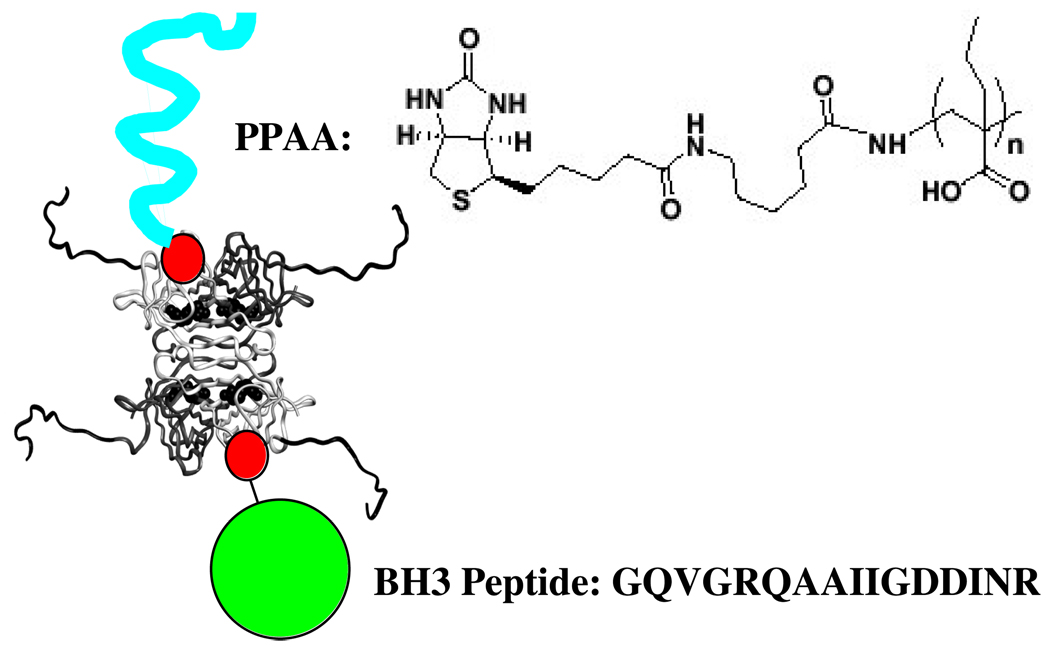
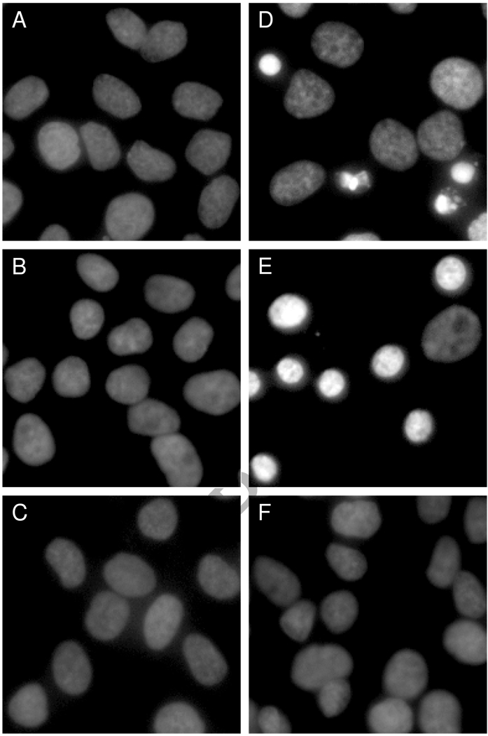
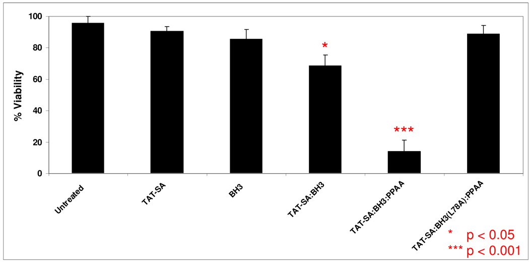
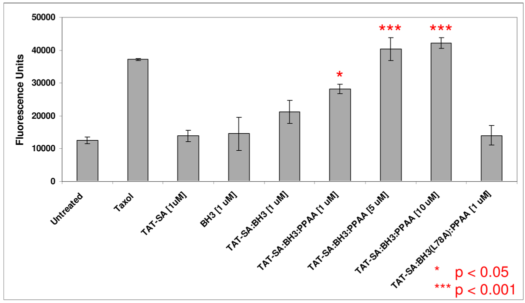
A key challenge in developing protein therapeutics or imaging agents that work against cytosolic targets is the intracellular delivery barrier. Here, we show that the pH-responsive, membrane-destabilizing polymer, poly (propylacrylic acid) (PPAA), can strongly enhance target cell killing through the intracellular delivery of a functional proapoptotic peptide. The Bak BH3 peptide induces apoptosis via antagonization of suppressor targets such as Bcl-2 and Bcl-x(L). A genetically-engineered streptavidin that contains an N-terminal TAT peptide sequence was used to optimize the pinocytotic cell uptake of biotinylated BH3 peptide and end-biotinylated PPAA. Fluorescence microscopic analysis of DAPI-stained HELA cells was used to quantitate apoptosis. Approximately 30% of cells treated with TAT-SA:BH3 complexes revealed morphologically distinct nuclear condensation, a hallmark of apoptosis. The incorporation of biotinylated PPAA had the effect of markedly enhancing the killing effect of BH3 peptides by an additional 55% (p<0.001) to a total cell killing efficiency of 85%. Caspase-3 activity was up-regulated in a TAT-SA:BH3:PPAA dose-dependent manner. The induction of apoptosis with the TAT-SA:BH3:PPAA complex was abrogated with the L78A BH3 peptide, that had been previously shown to knock-out antagonization activity. The caspase and L78A peptide results demonstrate that the delivered BH3 is indeed working through the biologically relevant apoptosis signaling pathway. These studies establish the ability of PPAA to strongly enhance the intracellular delivery of a functional pro-apoptotic peptide. Together with the PPAA, the TAT-SA adaptor complex could prove useful as a carrier of peptide/protein cargo to cultured cells.


Figures

Figure 1
Figure 1
Schematic representation of the TAT-SA complex with biotinylated PPAA and biotinylated BH3 peptide (biotin represented as red circle). The biotin linker and PPAA sequence are shown, together with the BH3 peptide sequence. The TAT peptide sequence has been modeled as a random coil extension onto the N-terminus of the four streptavidin subunits whose backbone folds are defined from x-ray crystallographic structures. They appear as the backbone extensions coming off the four subunits.
Figure 2
Figure 2
TAT-SA-mediated delivery of BH3 peptides induces apoptotic nuclear morphology in HeLa cells. Cells were treated with the indicated reagents at 10 µM for 24 h in serum-free DMEM before fluorescence microscopic analysis of DAPI-stained nuclei. Samples were A) untreated, B) TAT-SA, C) BH3, D) TAT-SA:BH3 1:1, E) TAT-SA: BH3:PPAA 1:1:1 and F) TAT-SA:BH3-(L78A):PPAA 1:1:1. The mutant BH3-(L78A) peptide, which is deficient in binding to Bcl-2-like proteins, was used as a negative control.
Figure 3
Figure 3
Quantitation of HeLa cell viability in response to TAT-SA-mediated delivery of BH3 peptides. Cells were treated with reagents at 10 µM for 24 h before fluorescence microscopy of DAPI-stained nuclei. The cell viability was defined as live or dead based on the presence or absence of condensed nuclear morphology visualized by the DAPI staining. Treatment of HeLa cells with TAT-SA:BH3 complexes resulted in an approximate 30% loss in cell viability (p<0.05, Tukey-Kramer) after 24 hours as compared to BH3-treated cells. The relative number of apoptotic to viable cells was markedly increased to approximately 85% (p<0.001, Tukey-Kramer) when the endosomal disruptive polymer PPAA was co-complexed to TAT-SA and BH3, but not when cells were treated with PPAA alone.
Figure 4
Figure 4
TAT-SA-mediated delivery of BH3 induces upregulation of caspase-3 activity in HeLa cells. Cells were treated with the indicated reagents for 16 hours in serum-free DMEM and their lysates subsequently assayed for caspase-3-mediated cleavage of a fluorescent substrate. The microtubule-damaging agent paclitaxel (Taxol) was used as a positive control at a concentration of 0.1 µM. Cells treated with TAT-SA:BH3 displayed slightly elevated caspase-3 activity compared to cells treated with TAT-SA alone or BH3 alone (p<0.05, Tukey-Kramer), demonstrating the ability of the BH3 peptide to induce apoptosis after the enhanced uptake of TAT-SA. The co-complexation of the endosomal releasing polymer PPAA resulted in increased caspase-3 activity in a dose-dependent fashion. Notably, the level of caspase-3 activity at the 5 µM and 10 µM conditions with PPAA was not statistically different (p>0.05, Tukey-Kramer), suggesting a saturation in the level of caspase-3 activity. Furthermore, when the mutant BH3-(L78A) peptide was complexed to TAT-SA, caspase-3 activity remained at basal levels, confirming the presence of a precise intracellular target for BH3 in order to induced apoptosis.

Similar articles

Cited by

References

    1. Fawell S, Seery J, Daikh Y, Moore C, Chen LL, Pepinsky B, Barsoum J. Tat-mediated delivery of heterologous proteins into cells. Proc. Natl. Acad. Sci. USA. 1994;91:664–668. - PMC - PubMed
    1. Derossi D, Joliot AH, Chassaing G, Prochiantz A. The third helix of the Antennapedia homeodomain translocates through biological membranes. J Biol Chem. 1994;269:10444–10450. - PubMed
    1. Elliott G, O'Hare P. Intercellular trafficking and protein delivery by a herpesvirus structural protein. Cell. 1997;88:223–233. - PubMed
    1. Schwarze SR, Dowdy SF. In vivo protein transduction: intracellular delivery of biologically active proteins, compounds and DNA. Trends Pharmacol. Sci. 2000;21:45–48. - PubMed
    1. Snyder EL, Dowdy SF. Protein/peptide transduction domains: potential to deliver large DNA molecules into cells. Curr. Opin. Mol. Ther. 2001;3:147–1452. - PubMed

LinkOut - more resources