

Achievement of three year remission with bevacizumab and irinotecan in recurrent glioblastoma multiforme: a case report
- PMID: 21499558
- PMCID: PMC3076043
- DOI: 10.4137/CMO.S6525
Achievement of three year remission with bevacizumab and irinotecan in recurrent glioblastoma multiforme: a case report
Abstract
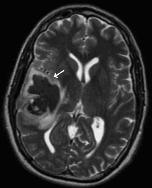
A 34-year-old man presented to the hospital with right-sided headache. He was diagnosed with GBM. He underwent resection of the tumor with placement of carmustine impregnated wafers. Then he underwent adjuvant chemotherapy with temozolamide. Before the completion of chemotherapy he had a recurrence. He underwent re-resection with placement of carmustine impregnated wafers. Subsequently he had eighteen cycles of salvage biochemotherapy with bevacizumab and irinotecan. To date, routine MRI scans of the brain have not shown evidence of recurrence. He continues to be in remission three years after treatment with bevacizumab and irinotecan.
Keywords: bevacizumab; irinotecan; recurrent glioblastoma multiforme.
Figures


Similar articles
-
Cerebrospinal fluid leak during treatment with bevacizumab and irinotecan after carmustine-impregnated wafers placement in patients with grade 2 oligodendroglioma and glioblastoma multiforme: report of two cases and review of literature.Cancer Invest. 2010 Dec;28(10):1048-53. doi: 10.3109/07357907.2010.483499. Epub 2010 Sep 27. Cancer Invest. 2010. PMID: 20873990 Review.
-
Reinduction of bevacizumab in combination with pegylated liposomal Doxorubicin in a patient with recurrent glioblastoma multiforme who progressed on bevacizumab/irinotecan.J Oncol. 2008;2008:942618. doi: 10.1155/2008/942618. Epub 2008 Sep 2. J Oncol. 2008. PMID: 19259336 Free PMC article.
-
Cs-131 brachytherapy for patients with recurrent glioblastoma combined with bevacizumab avoids radiation necrosis while maintaining local control.Brachytherapy. 2020 Sep-Oct;19(5):705-712. doi: 10.1016/j.brachy.2020.06.013. Brachytherapy. 2020. PMID: 32928486
-
Extensive Leptomeningeal Intracranial and Spinal Metastases in a Patient with a Supratentorial Glioblastoma Multiforme, IDH-Wildtype.World Neurosurg. 2018 Dec;120:442-447. doi: 10.1016/j.wneu.2018.09.082. Epub 2018 Sep 22. World Neurosurg. 2018. PMID: 30253992
-
Bevacizumab: a treatment option for recurrent glioblastoma multiforme.Ann Pharmacother. 2008 Oct;42(10):1486-90. doi: 10.1345/aph.1L030. Epub 2008 Sep 2. Ann Pharmacother. 2008. PMID: 18765835 Review.
Cited by
-
Patterns of long-term survivorship following bevacizumab treatment for recurrent glioma: a case series.CNS Oncol. 2019 Jun 1;8(2):CNS35. doi: 10.2217/cns-2019-0007. Epub 2019 Jul 11. CNS Oncol. 2019. PMID: 31293169 Free PMC article.
-
The role of brain tumor advocacy groups.Curr Neurol Neurosci Rep. 2014 Apr;14(4):442. doi: 10.1007/s11910-014-0442-z. Curr Neurol Neurosci Rep. 2014. PMID: 24604060 Review.
References
-
- DeAngelis LM. Brain tumors. N Engl J Med. 2001;344:114–23. - PubMed
-
- Jemal A, Siegel R, Ward E, et al. Cancer statistics, 2008. CA Cancer J Clin. 2008;58:71–96. - PubMed
-
- Buckner JC. Factors influencing survival in high-grade gliomas. Semin Oncol. 2003;30:10–4. - PubMed
-
- Balmaceda C, Peereboom D, Pannullo S, et al. Multi-institutional phase II study of temozolomide administered twice daily in the treatment of recurrent high-grade gliomas. Cancer. 2008;112:1139–46. - PubMed
-
- Wong ET, Hess KR, Gleason MJ, et al. Outcomes and prognostic factors in recurrent glioma patients enrolled onto phase II clinical trials. J Clin Oncol. 1999;17:2572–8. - PubMed
Publication types
LinkOut - more resources
Full Text Sources
