

Modulation of ammonium perfluorooctanoate-induced hepatic damage by genetically different PPARα in mice
- PMID: 21499893
- PMCID: PMC6594146
- DOI: 10.1007/s00204-011-0704-3
Modulation of ammonium perfluorooctanoate-induced hepatic damage by genetically different PPARα in mice
Abstract
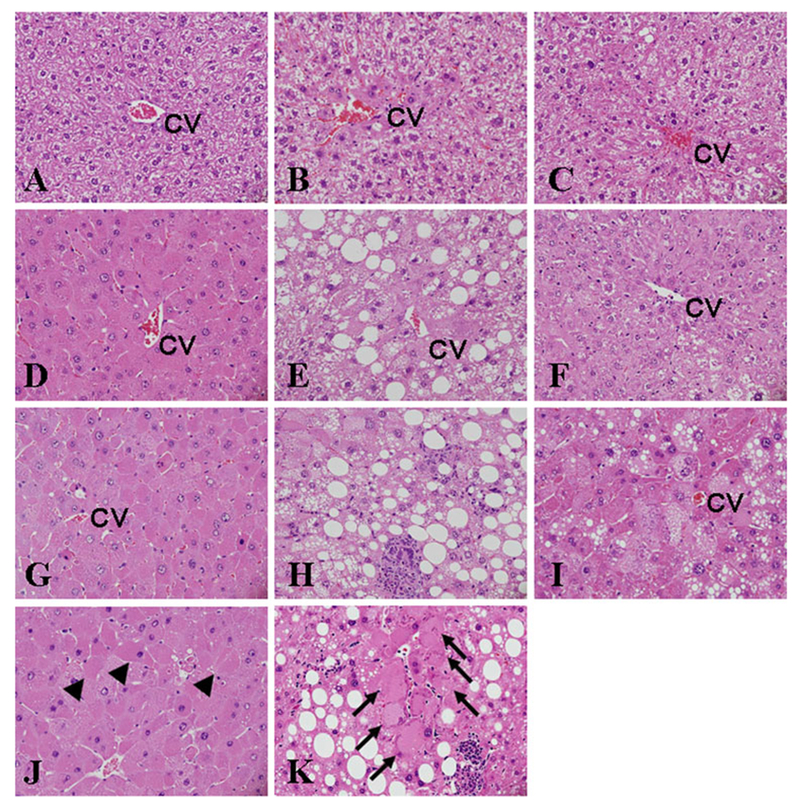
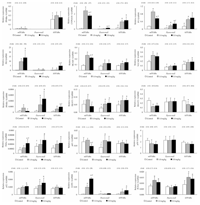
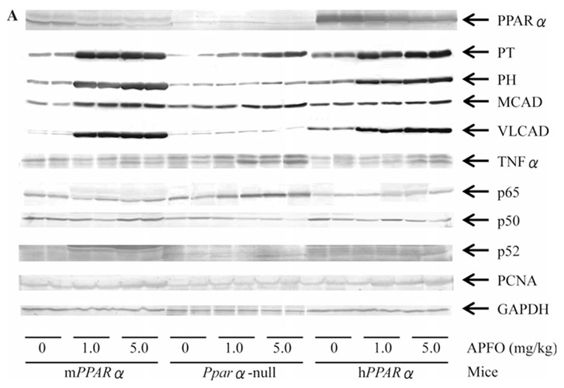
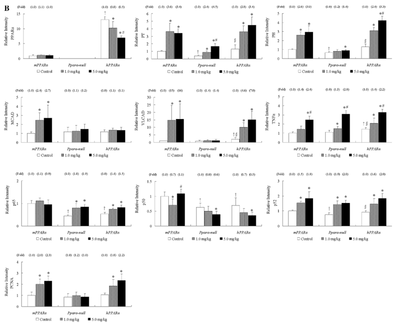
Perfluorooctanoic acid is a ligand for peroxisome proliferator-activated receptor (PPARα). Ammonium perfluorooctanoate (APFO) at 0.1 and 0.3 mg/kg doses activated mouse PPARα, but not human PPARα. This study aimed to clarify whether milligram-order APFO can activate human PPARα, and the receptor is involved in APFO-induced chronic hepatic damage. Male Sv/129 wild-type (mPPARα), Pparα-null, and humanized PPARα (hPPARα) mice (8 weeks old) were divided into three groups. The first was treated with water and the other two with 1.0 and 5.0 mg/kg APFO for 6 weeks, orally, respectively. Both doses activated mouse and human PPARα to a similar or lower degree in the latter. APFO dose dependently increased hepatic triglyceride levels in Pparα-null and hPPARα mice, but conversely decreased those in mPPARα ones. APFO-induced hepatic damage differed markedly among the three genotyped groups: single-cell necrosis was observed in all genotyped mice; inflammatory cells and macrovesicular steatosis only in Pparα-null mice; and microvesicular steatosis and hydropic degenerations in hPPARα and Pparα-null mice. The molecular mechanism underlying these differences may be attributable to those of gene expressions involved in lipid homeostasis (PPARα, β- and ω-oxidation enzymes, and diacylglycerol acyltransferases) and uncoupling protein 2. Thus, milligram-order APFO activated both mouse and human PPARα in a different manner, which may reflect histopathologically different types of hepatic damage.
Conflict of interest statement
Figures
Similar articles
-
Microgram-order ammonium perfluorooctanoate may activate mouse peroxisome proliferator-activated receptor alpha, but not human PPARalpha.Toxicology. 2009 Nov 9;265(1-2):27-33. doi: 10.1016/j.tox.2009.09.004. Epub 2009 Sep 12. Toxicology. 2009. PMID: 19751795 Free PMC article.
-
Ammonium perfluorooctanoate may cause testosterone reduction by adversely affecting testis in relation to PPARα.Toxicol Lett. 2011 Sep 10;205(3):265-72. doi: 10.1016/j.toxlet.2011.06.015. Epub 2011 Jun 25. Toxicol Lett. 2011. PMID: 21712084 Free PMC article.
-
Hepatic peroxisome proliferator-activated receptor α may have an important role in the toxic effects of di(2-ethylhexyl)phthalate on offspring of mice.Toxicology. 2011 Oct 28;289(1):1-10. doi: 10.1016/j.tox.2011.02.007. Epub 2011 Feb 24. Toxicology. 2011. PMID: 21354252
-
The role and regulation of the peroxisome proliferator activated receptor alpha in human liver.Biochimie. 2017 May;136:75-84. doi: 10.1016/j.biochi.2016.12.019. Epub 2017 Jan 8. Biochimie. 2017. PMID: 28077274 Review.
-
PPARalpha: mechanism of species differences and hepatocarcinogenesis of peroxisome proliferators.Toxicology. 2008 Apr 3;246(1):2-8. doi: 10.1016/j.tox.2007.09.030. Epub 2007 Oct 7. Toxicology. 2008. PMID: 18006136 Review.
Cited by
-
Hypercholesterolemia with consumption of PFOA-laced Western diets is dependent on strain and sex of mice.Toxicol Rep. 2016;3:46-54. doi: 10.1016/j.toxrep.2015.11.004. Toxicol Rep. 2016. PMID: 26942110 Free PMC article.
-
Risk to human health related to the presence of perfluorooctane sulfonic acid and perfluorooctanoic acid in food.EFSA J. 2018 Dec 13;16(12):e05194. doi: 10.2903/j.efsa.2018.5194. eCollection 2018 Dec. EFSA J. 2018. PMID: 32625773 Free PMC article.
-
Diet as an Exposure Source and Mediator of Per- and Polyfluoroalkyl Substance (PFAS) Toxicity.Front Toxicol. 2020 Dec 4;2:601149. doi: 10.3389/ftox.2020.601149. eCollection 2020. Front Toxicol. 2020. PMID: 35296120 Free PMC article. Review.
-
The EU's Per- and Polyfluoroalkyl Substances (PFAS) Ban: A Case of Policy over Science.Toxics. 2023 Aug 22;11(9):721. doi: 10.3390/toxics11090721. Toxics. 2023. PMID: 37755732 Free PMC article. Review.
-
Perfluorooctanoic acid increases serum cholesterol in a PPARα-dependent manner in female mice.Arch Toxicol. 2025 May;99(5):2087-2105. doi: 10.1007/s00204-025-03984-7. Epub 2025 Mar 1. Arch Toxicol. 2025. PMID: 40021516
References
-
- Aoyama T, Peters JM, Iritani N, Nakajima T, Furihata K, Hashimoto T, Gonzalez FJ (1998) Altered constitutive expression of fatty acid-metabolizing enzymes in mice lacking the peroxisome proliferator-activated receptor α (PPARα). J Biol Chem 273:5678–5684 - PubMed
-
- Berthiaume J, Wallace KB (2002) Perfluorooctanoate, perflourooc-tanesulfonate, and N-ethyl perfluorooctanesulfonamido ethanol: peroxisome proliferation and mitochondrial biogenesis. Toxicol Lett 129:23–32 - PubMed
-
- Biegel LB, Hurtt ME, Frame SR, O’Connor JC, Cook JC (2001) Mechanisms of extrahepatic tumor induction by peroxisome proliferators in male CD rats. Toxicol Sci 60:44–55 - PubMed
-
- Brunt EM, Janney CG, Di Bisceglie AM, Neuschwander-Tetri BA, Bacon BR (1999) Nonalcoholic steatohepatitis: a proposal for grading and staging the histological lesions. Am J Gastroenterol 94:2467–2474 - PubMed
-
- Butenhoff J, Costa G, Elcombe C, Farrar D, Hansen K, Iwai H, Jung R, Kennedy G Jr, Lieder P, Olsen G, Thomford P (2002) Toxicity of ammonium perfluorooctanoate in male cynomolgus monkeys after oral dosing for 6 months. Toxicol Sci 69:244–257 - PubMed
Publication types
MeSH terms
Substances
Grants and funding
LinkOut - more resources
Full Text Sources
Medical
