. 2011 May;36(5):404-16.
doi: 10.3109/02713683.2011.566411.

Functional characterization of folate transport proteins in Staten's Seruminstitut rabbit corneal epithelial cell line

Affiliations

Functional characterization of folate transport proteins in Staten's Seruminstitut rabbit corneal epithelial cell line

J Jwala et al. Curr Eye Res. 2011 May.

Abstract

Purpose: The overall objective of this study was to investigate and characterize the expression of folate transport proteins in Staten's Seruminstitut rabbit corneal (SIRC) epithelial cell line.

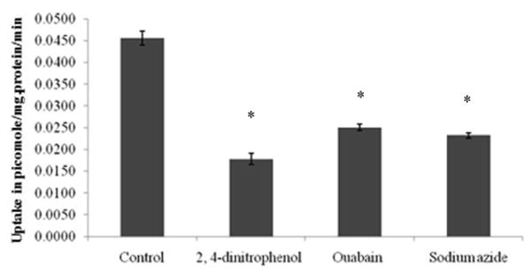
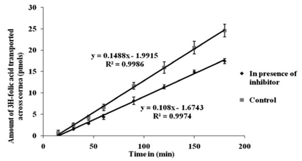
Methods: [(3)H]Folic acid uptake was studied with respect to time, pH, temperature, sodium, and chloride ion dependency. Inhibition studies were conducted with structural analogs, vitamins, and metabolic inhibitors. [(3)H]Folic acid uptake was also determined with varying concentrations of cold folic acid. Uptake kinetics was studied in the presence of various modulators of intracellular regulatory pathways, protein kinases A and C (PKA and PKC), protein tyrosine kinase (PTK), and calcium-calmodulin modulators. Ex vivo corneal permeability studies were carried out with [(3)H]folic acid in the presence and absence of 1 mM cold folic acid.

Results: Linear increase in [(3)H]folic acid uptake was observed over 30 min. The process followed saturation kinetics with apparent K(m) of 14.2 ± 0.2 nM, V(max) of (1.5 ± 0.1)*10(-5) micro.moles/min/mg protein, and K(d) of (2.1 ± 0.2)*10(-6) min(-1). The uptake process was found to be dependent on pH, sodium ions, chloride ions, temperature, and energy. Uptake was inhibited in the presence of structural analogs (cold folic acid, methyltetrahydro folate, and methotrexate), but structurally unrelated vitamins did not show any effect. Membrane transport inhibitors SITS, DIDS, probenecid and endocytic inhibitor, colchicine significantly inhibited the [(3)H]folic acid uptake indicating the involvement of receptor/transporter mediated process. PKA, PTK, and Ca(2+)/calmodulin pathways significantly regulate the process. RT-PCR and Western blot analysis confirmed the presence of folate receptor-α (FR-alpha) and proton-coupled folate transporter (PCFT). Permeability of [(3)H]folic acid across the rabbit cornea was (1.48 ± 0.13)*10(-05) cm/sec, and in the presence of cold folic acid it was (1.08 ± 0.10)*10(-05) cm/sec.

Conclusions: This work demonstrated the functional and molecular presence of FR-alpha and PCFT in SIRC epithelial cell line.


Figures

Fig. 1
Fig. 1
Uptake of [3H]Folic acid by SIRC cells as a function of time. Each data point represents the mean α standard deviation of 5 separate uptake determinations.
Fig. 2
Fig. 2
Uptake of [3H]Folic acid by SIRC cells as a function of pH. Each data point represents the mean α standard deviation of 5 separate uptake determinations. Asterisk (*) represents significant difference from control (p < 0.05).
Fig. 3
Fig. 3
Uptake of [3H]Folic acid by SIRC cells as a function of temperature. Each data point represents the mean α standard deviation of 5 separate uptake determinations. Asterisk (*) represents significant difference from control (p < 0.05).
Fig. 4
Fig. 4
Uptake of [3H]Folic acid by SIRC cells as a function of ions. Each data point represents the mean α standard deviation of 5 separate uptake determinations. Asterisk (*) represents significant difference from control (p < 0.05).
Fig. 5
Fig. 5
Uptake of [3H]Folic acid by SIRC cells in presence of various energy inhibitors. Each data point represents the mean α standard deviation of 5 separate uptake determinations. Asterisk (*) represents significant difference from control (p < 0.05).
Fig. 6
Fig. 6
Uptake of [3H]Folic acid by SIRC in presence of membrane transport inhibitors and endocytosis inhibitor colchicine. Each data point represents the mean α standard deviation of 5 separate uptake determinations. Asterisk (*) represents significant difference from control (p < 0.05).
Fig. 7
Fig. 7
Substrate specificity of uptake of [3H]Folic acid (10 nM) by SIRC cells in presence of various structurally related and unrelated analogs. Each data point represents the mean standard deviation of 5 separate uptake determinations. Asterisk (*) represents significant difference from control (p < 0.05).
Fig. 8
Fig. 8
Uptake of [3H]Folic acid (10 nM) in presence of various concentrations of cold folic acid on SIRC cell line.Saturation kinetic parameters are as follows : Vmax: 1.5*10-5, Km : 14.22 nM and Kd: 2.1*10-6 min.-1 Each data point represents the mean standard deviation of 5 separate uptake determinations.
Fig. 9
Fig. 9
Effect of Ca2+/calmodulin-mediated pathways modulators on the uptake of [3H]Folic acid in SIRC cells. Each data point represents the mean standard deviation of 5 separate uptake determinations. Asterisk (*) represents significant difference from control (p < 0.05).
Fig. 10
Fig. 10
Effect of PKA pathway modulators on the uptake of [3H]Folic acid in SIRC cells. Each data point represents the mean standard deviation of 5 separate uptake determinations. Asterisk (*) represents significant difference from control (p < 0.05).
Fig. 11
Fig. 11
Effect of PTK pathway modulators on the uptake of [3H]Folic acid in SIRC cells. Each data point represents the mean standard deviation of 5 separate uptake determinations. Asterisk (*) represents significant difference from control (p < 0.05).
Fig. 12
Fig. 12
RT-PCR analysis of FR-α (Folate receptor), RFC (reduce folate carrier), PCFT (Proton coupled folate transporter). GAPDH (Glyceraldehyde 3-phosphate dehydrogenase)
Fig. 13
Fig. 13
Western blot analysis of FR-α (Folate receptor) and PCFT (Proton coupled folate transporter).
Fig. 14
Fig. 14
Ex vivo permeability of [3H]Folic acid in excised rabbit cornea. Each data point represents the mean standard deviation of 4 separate determinations.

References

    1. Bozard BR, Ganapathy PS, Duplantier J, et al. Molecular and biochemical characterization of folate transport proteins in retinal Muller cells. Investigative ophthalmology & visual science. 51:3226–3235. - PMC - PubMed
    1. Brzezinska A, Winska P, Balinska M. Cellular aspects of folate and antifolate membrane transport. Acta Biochim Pol. 2000;47:735–749. - PubMed
    1. Golnik KC, Schaible ER. Folate-responsive optic neuropathy. J Neuroophthalmol. 1994;14:163–169. - PubMed
    1. Matherly LH, Goldman DI. Membrane transport of folates. Vitam Horm. 2003;66:403–456. - PubMed
    1. Sierra EE, Goldman ID. Recent advances in the understanding of the mechanism of membrane transport of folates and antifolates. Semin Oncol. 1999;26:11–23. - PubMed

Publication types

MeSH terms

Substances

LinkOut - more resources