. 2011 Oct;10(5):769-79.
doi: 10.1111/j.1474-9726.2011.00714.x. Epub 2011 May 25.

MicroRNA-18 and microRNA-19 regulate CTGF and TSP-1 expression in age-related heart failure

Affiliations

MicroRNA-18 and microRNA-19 regulate CTGF and TSP-1 expression in age-related heart failure

Geert C van Almen et al. Aging Cell. 2011 Oct.

Abstract

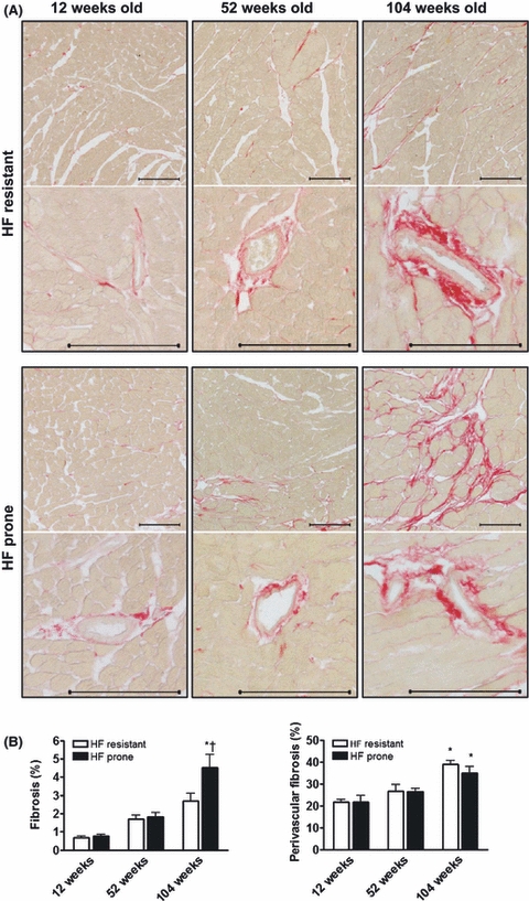
To understand the process of cardiac aging, it is of crucial importance to gain insight into the age-related changes in gene expression in the senescent failing heart. Age-related cardiac remodeling is known to be accompanied by changes in extracellular matrix (ECM) gene and protein levels. Small noncoding microRNAs regulate gene expression in cardiac development and disease and have been implicated in the aging process and in the regulation of ECM proteins. However, their role in age-related cardiac remodeling and heart failure is unknown. In this study, we investigated the aging-associated microRNA cluster 17-92, which targets the ECM proteins connective tissue growth factor (CTGF) and thrombospondin-1 (TSP-1). We employed aged mice with a failure-resistant (C57Bl6) and failure-prone (C57Bl6 × 129Sv) genetic background and extrapolated our findings to human age-associated heart failure. In aging-associated heart failure, we linked an aging-induced increase in the ECM proteins CTGF and TSP-1 to a decreased expression of their targeting microRNAs 18a, 19a, and 19b, all members of the miR-17-92 cluster. Failure-resistant mice showed an opposite expression pattern for both the ECM proteins and the microRNAs. We showed that these expression changes are specific for cardiomyocytes and are absent in cardiac fibroblasts. In cardiomyocytes, modulation of miR-18/19 changes the levels of ECM proteins CTGF and TSP-1 and collagens type 1 and 3. Together, our data support a role for cardiomyocyte-derived miR-18/19 during cardiac aging, in the fine-tuning of cardiac ECM protein levels. During aging, decreased miR-18/19 and increased CTGF and TSP-1 levels identify the failure-prone heart.


Figures

Fig. 1
Fig. 1
Enhanced left ventricular interstitial fibrosis in old heart failure (HF) prone mice. (A) Histological analysis of the hearts of HF-resistant and HF-prone mice by Sirius Red staining. Photographs show representative areas of interstitial fibrosis (upper panel) and collagen deposition in the perivascular area (lower panel). Scale bars represent 100 μm. (B) Quantitative analysis of the interstitial and perivascular collagen content in HF-resistant (12 weeks, n = 8; 52 weeks, n = 8; and 104 weeks, n = 9) and HF-prone mice (12 weeks, n = 6; 52 weeks, n = 11; and 104 weeks, n = 9) revealed increased interstitial fibrosis in the left ventricles of 104-week-old HF-prone mice. Perivascular fibrosis was significantly increased in 104-week-old hearts, but was not different between HF-resistant and HF-prone mice. Data are presented as mean ± SEM. *P≤0.05 vs 52-week-old HF-prone mice; †P≤0.05 vs 104-week-old HF-resistant mice.
Fig. 2
Fig. 2
Opposite expression profiles in heart failure (HF)-resistant versus HF-prone mice. CTGF, TSP-1, miR-18a, miR-19a, and miR-19b levels in aged HF-resistant (12 weeks, n = 8; 52 weeks, n = 8; and 104 weeks, n = 9) and HF-prone mice (12 weeks, n = 6; 52 weeks, n = 11; and 104 weeks, n = 9). (A and B) Immunoblotting was performed on four mice per age-group and revealed significant induction of CTGF and TSP-1 protein expression in failing hearts of 104-week-old HF-prone mice, whereas CTGF and TSP-1 levels were reduced in old HF-resistant mice. Immunoblots show representative protein bands of TSP-1, CTGF, and GAPDH. (C) RT-PCR analysis showed increased expression of miR-18a, miR-19a, and miR-19b in 104-week-old HF-resistant hearts, whereas age-matched HF-prone mice had decreased expressions. miRNA expression and CTGF and TSP-1 protein levels were normalized for GAPDH expression and presented as mean ± SEM. *P≤0.05 vs 12 weeks of age; †P≤0.05 vs 52 weeks of age; $P = 0.05 vs 12 weeks of age.
Fig. 3
Fig. 3
CTGF and TSP-1 expression are elevated in human heart failure (HF). RT-PCR analysis of miR-18a, miR-19a, miR-19b, CTGF, and TSP-1 transcript levels in myocardial biopsies from idiopathic cardiomyopathy (ICM) patients at older age with normal (n = 5) and severely impaired (n = 9) cardiac function. Transcript levels were compared to the expression in young ICM subjects with a preserved cardiac function (n = 5). (A) MiR-18a, miR-19a, and miR-19b expression was significantly decreased in older ICM patients with HF. (B) CTGF and TSP-1 transcript levels were significantly induced in older patients with a compromised cardiac function, when compared to younger ICM subjects. All data were normalized for GAPDH expression and presented as mean ± SEM. *P≤0.05 vs young ICM patients. $Pa = 0.06 in failing vs nonfailing hearts of older ICM patients.
Fig. 4
Fig. 4
Aging-induced expression profiles in cardiomyocytes in vitro. (A) Electron microscopic images of the left ventricle of 12- and 104-week-old mice, and 4- and 21-day-old neonatal rat cardiomyocytes (NRCMs) showing perinuclear accumulation of lipofuscin in cardiomyocytes of 104-week-old mice and NRCMs after 21 days in culture. Scale bars represent 2 μm. (B) Two-photon/confocal images and quantification of autofluorescent lipofuscin granules (green) in cultured cardiomyocytes. Nuclei are stained with Hoechst (blue). Scale bars represent 100 μm. (C) RT-PCR analysis showed significant induction of collagen type 3A1 (COL3A1), but not collagen type 1A1 (COL1A1) in cultured NRCMs. (D) RT-PCR analysis revealed decreased miR-18a, miR-19a, and miR-19b expression in aged NRCMs after 21 days in culture. (E) CTGF and TSP-1 transcript levels increased with NRCM aging in vitro. (F) Immunoblotting confirmed the increase in CTGF and TSP-1 protein induction during cardiomyocyte aging. All in vitro experiments were performed with n = 3 per group, and protein and transcript levels were normalized for GAPDH expression. Data were presented as mean ± SEM. *P≤0.05 vs 4-day-old NRCMs.
Fig. 5
Fig. 5
MiR-18a and miR-19b regulate CTGF and TSP-1 expression in cardiomyocytes. (A–C) In situ hybridization showed the abundant expression of miR-18a and miR-19b in the myocardium of adult C57Bl6 mice, most of it expressed by cardiomyocytes. Black arrows indicate the cardiomyocyte nucleus and illustrate the perinuclear localization of these miRNAs. (D and E) Comparison of the expression profiles in cultured neonatal rat cardiomyocytes (NRCMs) and neonatal rat cardiac fibroblasts (NRCFs) shows that abundant expression of miR-18a and miR-19b in NRCMs is paralleled by low CTGF and TSP-1 transcript levels. (F and G) Immunoblotting revealed that manipulating miR-18a and miR-19b function by overexpression of these miRNAs using mimics in NRCMs was sufficient to decrease CTGF and TSP-1 protein expression, while inhibition with antagomirs enhanced CTGF and TSP-1 levels. (H and I) In contrast to NRCMs, immunoblotting in cultured NRCFs showed that CTGF and TSP-1 protein expression was not suppressed by overexpression of miR-18a and miR-19b, nor did inhibition of these miRNAs result in increased CTGF and TSP-1 levels. Mimic and antagomir experiments were performed with n = 4 per group, and data were normalized for GAPDH expression. Data were presented as mean ± SEM. *P≤0.05 vs scrambled control oligonucleotides.
Fig. 6
Fig. 6
MiR-18a and miR-19b regulate collagen 1A1 and 3A1 expression in cardiomyocytes in vitro. RT-PCR analysis for the induction collagen 1A1 (COL1A1) and collagen 3A1 (COL3A1) in cultured neonatal rat cardiomyocytes and cardiac fibroblasts after manipulation with miR-18a and miR-19b mimics and antagomirs. (A) Overexpression of miR-18a and miR-19b in cardiomyocytes significantly reduces collagen 1A1 and collagen 3A1 transcript levels, while inhibition of these miRNAs using antagomirs significantly induced transcription of both collagen types. (B) Collagen 1A1 and 3A1 expression in cultured cardiac fibroblasts seemed unrelated to miR-18a and miR-19b, as no significant repression or induction was observed in NRCFs after treatment with miR-18a and miR-19b mimics or antagomirs, respectively. All experiments were performed with n = 4 per group, and data were normalized for GAPDH transcript levels. Data were presented as mean ± SEM. *P≤0.05 vs scrambled control oligonucleotides.

References

    1. Barasch E, Gottdiener JS, Aurigemma G, Kitzman DW, Han J, Kop WJ, Tracy RP. Association between elevated fibrosis markers and heart failure in the elderly: the cardiovascular health study. Circ. Heart Fail. 2009;2:303–310. - PMC - PubMed
    1. Belmadani S, Bernal J, Wei CC, Pallero MA, Dell'italia L, Murphy-Ullrich JE, Berecek KH. A thrombospondin-1 antagonist of transforming growth factor-beta activation blocks cardiomyopathy in rats with diabetes and elevated angiotensin II. Am. J. Pathol. 2007;171:777–789. - PMC - PubMed
    1. Bodyak N, Kang PM, Hiromura M, Sulijoadikusumo I, Horikoshi N, Khrapko K, Usheva A. Gene expression profiling of the aging mouse cardiac myocytes. Nucleic Acids Res. 2002;30:3788–3794. - PMC - PubMed
    1. Boehm M, Slack F. A developmental timing microRNA and its target regulate life span in C. elegans. Science. 2005;310:1954–1957. - PubMed
    1. Boyle AJ, Shih H, Hwang J, Ye J, Lee B, Zhang Y, Kwon D, Jun K, Zheng D, Sievers R, Angeli F, Yeghiazarians Y, Lee R. Cardiomyopathy of aging in the mammalian heart is characterized by myocardial hypertrophy, fibrosis and a predisposition towards cardiomyocyte apoptosis and autophagy. Exp. Gerontol. 2011 in press. - PMC - PubMed

Publication types

MeSH terms

LinkOut - more resources