. 2011 Apr 18:11:144.
doi: 10.1186/1471-2407-11-144.

Curcumin-induced HDAC inhibition and attenuation of medulloblastoma growth in vitro and in vivo

Affiliations

Curcumin-induced HDAC inhibition and attenuation of medulloblastoma growth in vitro and in vivo

Seung Joon Lee et al. BMC Cancer. .

Abstract

Background: Medulloblastoma is the most common brain tumor in children, and its prognosis is worse than for many other common pediatric cancers. Survivors undergoing treatment suffer from serious therapy-related side effects. Thus, it is imperative to identify safer, effective treatments for medulloblastoma. In this study we evaluated the anti-cancer potential of curcumin in medulloblastoma by testing its ability to induce apoptosis and inhibit tumor growth in vitro and in vivo using established medulloblastoma models.

Methods: Using cultured medulloblastoma cells, tumor xenografts, and the Smo/Smo transgenic medulloblastoma mouse model, the antitumor effects of curcumin were tested in vitro and in vivo.

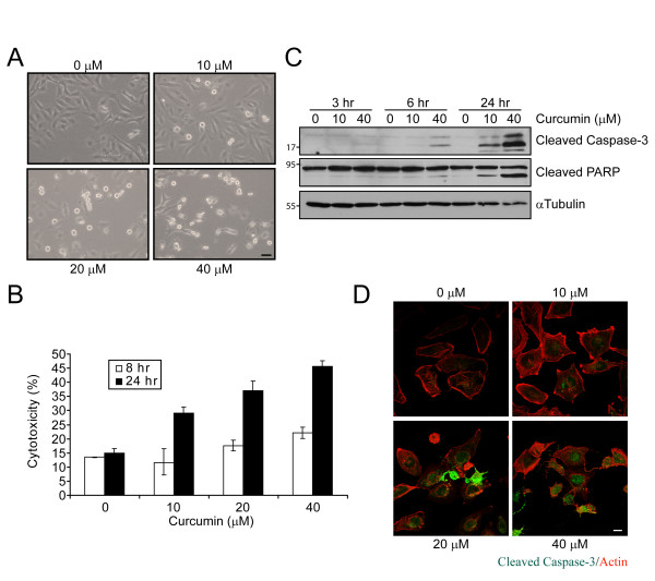
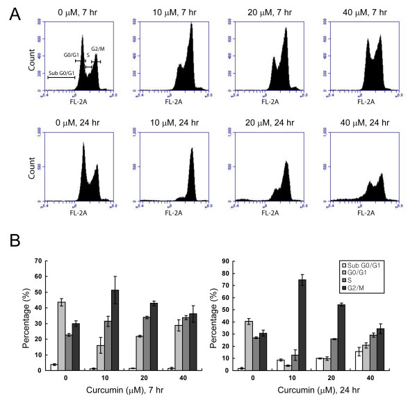
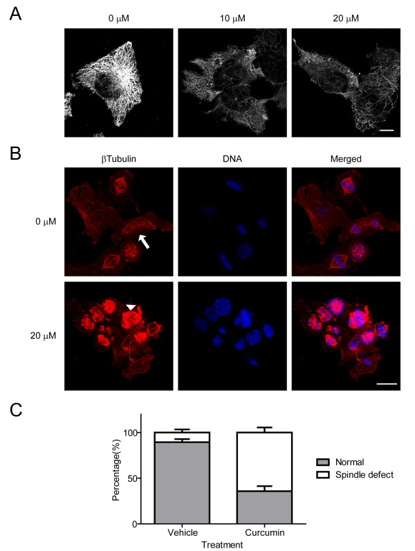
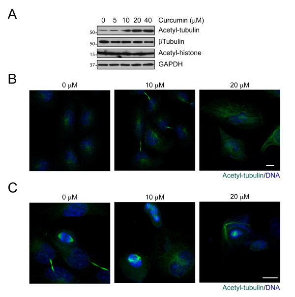
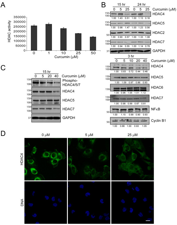
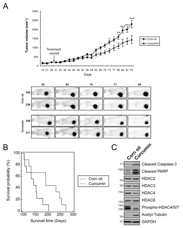
Results: Curcumin induced apoptosis and cell cycle arrest at the G2/M phase in medulloblastoma cells. These effects were accompanied by reduced histone deacetylase (HDAC) 4 expression and activity and increased tubulin acetylation, ultimately leading to mitotic catastrophe. In in vivo medulloblastoma xenografts, curcumin reduced tumor growth and significantly increased survival in the Smo/Smo transgenic medulloblastoma mouse model.

Conclusions: The in vitro and in vivo data suggest that curcumin has the potential to be developed as a therapeutic agent for medulloblastoma.


Figures

Figure 1
Figure 1
Curcumin induces apoptosis in medulloblastoma cells. A. Phase contrast images of DAOY cells incubated with indicated concentrations of curcumin for 16 hours. Bar, 100 μm. B. LDH release as measure of the cytotoxic effect of curcumin. C. Immunoblot of cleaved caspase-3 and PARP in curcumin-treated DAOY cells. α-tubulin immunoblot confirms that equal amounts of protein were used in the analysis. D. Immunofluorescence of control and curcumin-treated DAOY cells (16 hours) for cleaved capase-3 (green) and F-actin (red). Bar, 20 μm.
Figure 2
Figure 2
Curcumin elicits G2/M phase arrest. A. Cell cycle profiles of curcumin-incubated DAOY cells as analyzed by flow cytometry. Cells were exposed to curcumin at the indicated concentrations for 7 hours (top) and 24 hours (bottom). Note the increase in cells arrested in G2/M upon curcumin treatment. B. Quantitative analysis of cells in A obtained from three independent experiments. Averages of mean ± SD are shown.
Figure 3
Figure 3
Curcumin affects microtubule dynamics and mitotic spindle assembly. A. Immunofluorescence for β-tubulin in DAOY cells treated with DMSO (0 μM) or curcumin (10, 20 μM) for 6 hours. Bar, 20 μm. B. Analysis of mitotic spindle microtubules in DAOY cells. Mitotic cells were released from the G2/M arrest in the presence or absence of curcumin. The cells were fixed after 60 min of release and stained for β-tubulin (red) and DNA (blue). The arrow indicates segregation of chromosomes along the mitotic spindle, while the arrowhead shows abnormal spindle formation and missegregation of chromosomes. Bar, 20 μm. C. Quantitative analysis of cells with abnormal mitotic spindles after 60 min of incubation with either vehicle or 20 μM curcumin. Error bars indicate standard deviations of three independent experiments.
Figure 4
Figure 4
Curcumin induces tubulin acetylation. A. DAOY cells were exposed to increasing concentration of curcumin for 3 hours and then immunoblotted with antibodies indicated. Increased acetylation of tubulin was also observed in interphase (B) and mitotic (C) cells after 16 hours of curcumin treatment. Methanol-fixed cells were immunostained with antibody specific for acetylated tubulin (green). DNA stained blue. Bar, 20 μm.
Figure 5
Figure 5
Curcumin blocks HDAC activity. A. Curcumin-treated DAOY cells (6 hours) were lysed and subjected to a fluoremetric HDAC activity assay as described in Materials and Methods. Data represent the average of the mean ± SEM of two independent experiments done in triplicates B. C. Expression levels of various HDAC family members (B) and phosphorylation level of HDAC4/5/7 (C) in DAOY cells treated with curcumin for 3, 15 and 24 hours. Representative immunoblots are shown. GAPDH immunoblots indicate loading of equal amounts of protein. Numbers below immunoblots in B are the mean of the intensity of three independent experiments, normalized to control cells at each time point and concentration. D. Subcellular localization of HDAC4 (green) in DAOY cells. Incubation with curcumin (6 hours) did not significantly affect the cellular distribution of HDAC4. Nuclei are indicated by DNA (blue). Bar, 30 μm.
Figure 6
Figure 6
Curcumin reduces tumor growth in DAOY in vivo tumor xenografts and Smo/Smo mice. A. Effect of curcumin on tumor growth of subcutaneous xenograft tumors in nude mice. 30 days after cell injection (arrow), each group of mice (N = 12) received corn oil or curcumin (1 g/kg body weight) once daily and the tumor volume was measured by using a caliper. Data are expressed as mean ± SEM. *, P < 0.05; **, P < 0.01; ***, P < 0.001. Data are representative of three independent studies. Bottom panel, fluorescent ventral images of tumor-bearing mice. tdTomato-expressing tumors were imaged once a week and representative images are shown. B. Life span of control and curcumin-treated Smo/Smo (N = 9) plotted as Kaplan-Meier survival curve. The median survival time of curcumin-treated mice was 192 days vs 144 days of control mice (corn oil). P = 0.0308 (log rank analysis). C. Apoptotic markers, tubulin acetylation and HDAC expression and phosphorylation in tumor lysates obtained from each group of the Smo/Smo mice.

References

    1. Packer RJ. Childhood Brain Tumors: Accomplishments and Ongoing Challenges. J Child Neurol. 2008;23(10):1122–1127. doi: 10.1177/0883073808320758. - DOI - PMC - PubMed
    1. Gilbertson RJ, Ellison DW. The origins of medulloblastoma subtypes. Annu Rev Pathol. 2008;3:341–365. doi: 10.1146/annurev.pathmechdis.3.121806.151518. - DOI - PubMed
    1. Fossati P, Ricardi U, Orecchia R. Pediatric medulloblastoma: Toxicity of current treatment and potential role of protontherapy. Cancer Treatment Reviews. 2009;35(1):79–96. doi: 10.1016/j.ctrv.2008.09.002. - DOI - PubMed
    1. Schuller U, Heine VM, Mao J, Kho AT, Dillon AK, Han YG, Huillard E, Sun T, Ligon AH, Qian Y. et al.Acquisition of granule neuron precursor identity is a critical determinant of progenitor cell competence to form Shh-induced medulloblastoma. Cancer Cell. 2008;14(2):123–134. doi: 10.1016/j.ccr.2008.07.005. - DOI - PMC - PubMed
    1. Romer JT, Kimura H, Magdaleno S, Sasai K, Fuller C, Baines H, Connelly M, Stewart CF, Gould S, Rubin LL. et al.Suppression of the Shh pathway using a small molecule inhibitor eliminates medulloblastoma in Ptc1(+/-)p53(-/-) mice. Cancer Cell. 2004;6(3):229–240. doi: 10.1016/j.ccr.2004.08.019. - DOI - PubMed

Publication types

MeSH terms

LinkOut - more resources