. 2011 Jun;92(6):521-7.
doi: 10.1016/j.exer.2011.03.015. Epub 2011 Apr 9.

Delayed post-ischemic conditioning significantly improves the outcome after retinal ischemia

Affiliations

Delayed post-ischemic conditioning significantly improves the outcome after retinal ischemia

John C Dreixler et al. Exp Eye Res. 2011 Jun.

Abstract

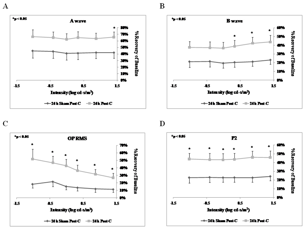
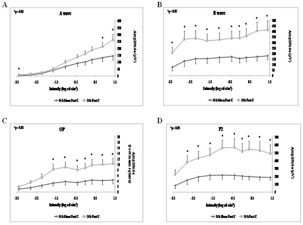
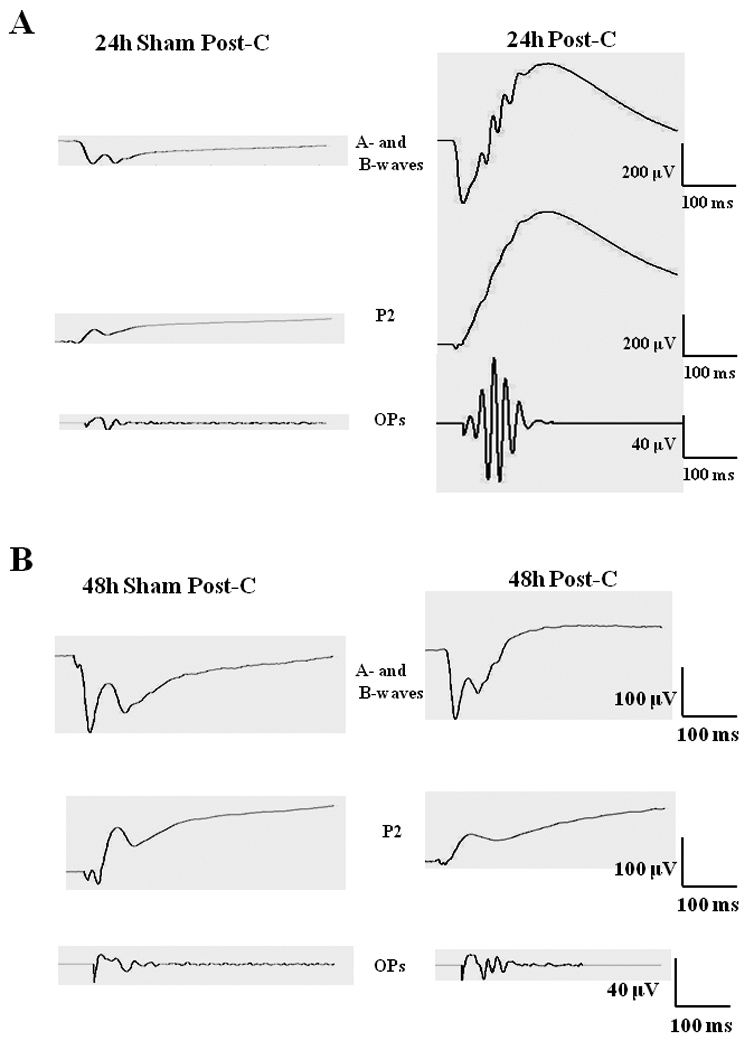
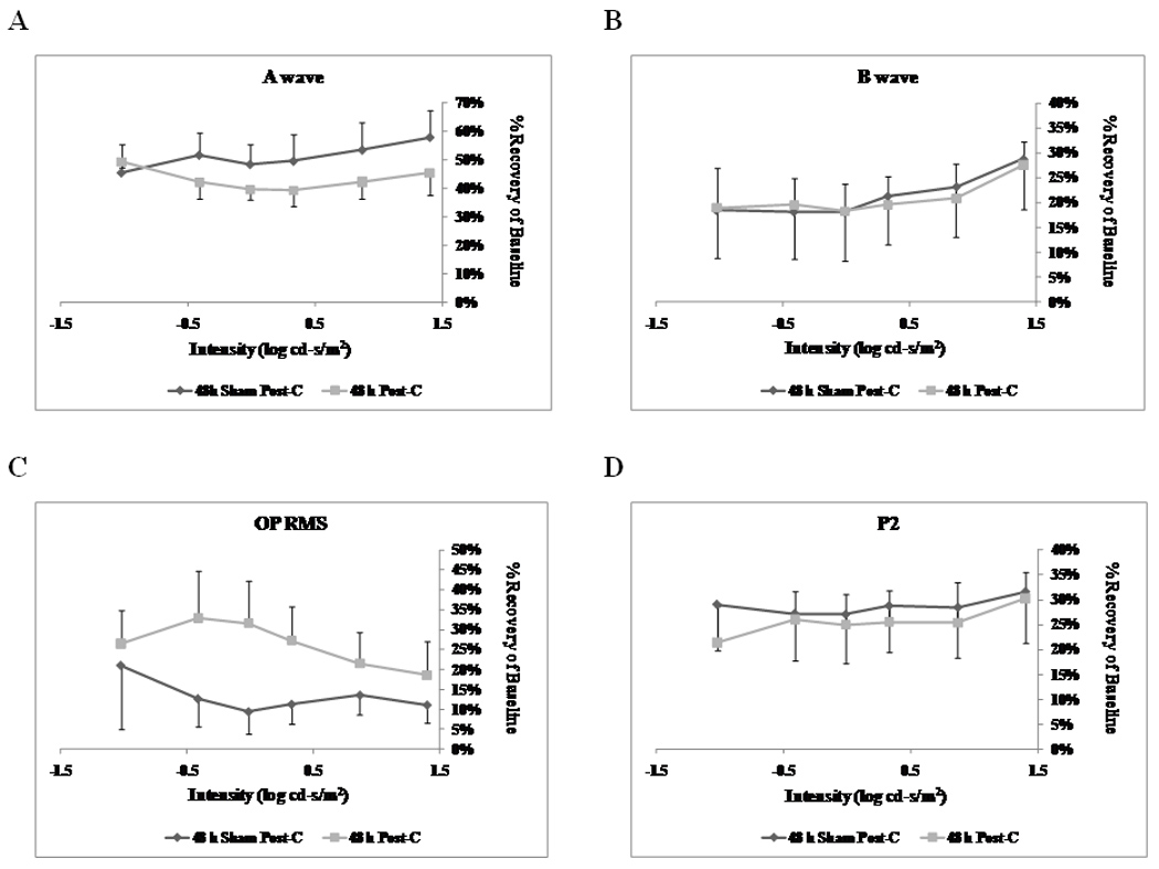
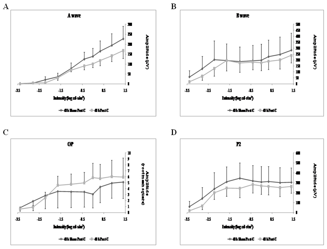
In previous studies, it was shown that post-conditioning, a transient period of brief ischemia following prolonged severe ischemia in the retina, could provide significant improvement in post-ischemic recovery, attenuation of cell loss, and decreased apoptosis. These studies showed that post-conditioning effectively prevented damage after retinal ischemia when it was instituted early (within 1 h) in the post-ischemic period. While post-ischemic conditioning holds high promise of clinical translation, patients often present late after the onset of retinal ischemia and therefore immediate application of this anti-ischemic maneuver is generally not feasible. In this study, we examined the hypothesis that application of a post-conditioning stimulus at 24 h or greater following the end of prolonged ischemia would decrease the extent of ischemic injury. Ischemia was induced in rat retina in vivo. Recovery after ischemia followed by 5 min of post-conditioning brief ischemia 24 or 48 h after prolonged ischemia was assessed functionally (electroretinography) and histologically at 7 days after ischemia and post-conditioning or sham post-conditioning. We found that the brief ischemic stimulus applied 24, but not 48 h after prolonged ischemia significantly improved functional recovery and decreased histological damage induced by prolonged ischemia. We conclude that within a defined time window, delayed post-ischemic conditioning ameliorated post-ischemic injury in rats. Compared to earlier studies, the present work demonstrates for the first time the novel ability of a significantly delayed ischemic stimulus to provide robust neuroprotection in the retina following ischemia.


Conflict of interest statement

There is no conflict of interest or commercial interest for any of the authors.

Figures

Figure 1
Figure 1
Stimulus-Intensity response in post-conditioned retinae for 24 h post-C vs 24 h sham post-C. Double normalized (corrected for the non-ischemic eye and for diurnal variation from baseline to 7 days after post-conditioning) ERG data for a-, b-wave, oscillatory potentials (sum of root mean square, OP RMS) and P2 over a range of flash intensities from −1.02 to 1.40 log cd-s/m2. The data were recorded at baseline (prior to ischemia) and at 7 days after post-conditioning; post-C was applied 24 h after ischemia. ERG data for a- (A), b-wave (B), OP RMS(C) and P2 (D) over a range of flash intensities suggests that 24 h delayed post-C was functionally protective. Solid lines with diamonds = 24 h sham post-C (n = 11). Solid lines with squares = 24 h post-C (n = 15).
Figure 2
Figure 2
Stimulus-Intensity response in post-conditioned retinae for 24 h post-C vs 24 h sham post-C. Absolute ERG data for a-, b-wave, oscillatory potentials (sum of root mean square, OP RMS) and P2 over a range of flash intensities from −1.02 to 1.40 log cd-s/m2. The data were recorded at baseline (prior to ischemia) and at 7 days after post- conditioning; post-C was applied 24 h after ischemia. ERG data for a- (A), b-wave (B), OP RMS(C) and P2 (D) over a range of flash intensities suggests that 24 h delayed post-C was functionally protective. Solid lines with diamonds = 24 h sham post-C (n = 11). Solid lines with squares = 24 h post-C (n = 15).
Figure 3
Figure 3
(A) Representative ERG traces at 0.87 log cd.s/m2 flash intensity for the a- and b-wave (top), P2 (middle) and the OPs (bottom) for both 24 h post-C and 24 h sham post-C groups. (B) Representative ERG traces at 0.87 log cd.s/m2 flash intensity for the a- and b-wave (top), P2 (middle) and the OPs (bottom) for both 48 h post-C and 48 h sham post-C groups.
Figure 4
Figure 4
Representative histopathological images of hematoxylin and eosin-stained retinae in 4 µm thick sections for each of the experimental groups. These sections were prepared from retinae removed from the rats at 7 days following delayed post-C. Arrows indicate layers demonstrating cell loss and asterisks denote regions of inflammatory cell infiltration. Below each representative retina is a close-up of the RGC layer.
Figure 5
Figure 5
Stimulus-Intensity response in post-conditioned retinae for 48 h post-C vs 48 h sham post-C. Double normalized (corrected for the non-ischemic eye and for diurnal variation from baseline to 7 days after post-C) ERG data for a-, b-wave, oscillatory potentials (sum of root mean square, OP RMS) and P2 over a range of flash intensities from −1.02 to 1.40 log cd-s/m2. The data were recorded at baseline (prior to ischemia) and at 7 days after post-conditioning; post-C was applied 48 h after ischemia. ERG data for a- (A), b-wave (B), OP RMS(C) and P2 (D) over a range of flash intensities suggests that 48 h delayed post-C was not functionally protective. Solid lines with diamonds = 48 h sham post-C (n = 4). Solid lines with squares = 48 h post-C (n = 4).
Figure 6
Figure 6
Stimulus-Intensity response in post-conditioned retinae for 48 h post-C vs 48 h sham post-C. Absolute ERG data for a-, b-wave, oscillatory potentials (sum of root mean square, OP RMS) and P2 over a range of flash intensities from −1.02 to 1.40 log cd-s/m2. The data were recorded at baseline (prior to ischemia) and at 7 days after post-conditioning; post-C was applied 48 h after ischemia. ERG data for a- (A), b-wave (B), OP RMS(C) and P2 (D) over a range of flash intensities suggests that 48 h delayed post-C was not functionally protective. Solid lines with diamonds = 48 h sham post-C (n = 4). Solid lines with squares = 48 h post-C (n = 4).

References

    1. Bui BV, Edmunds B, Cioffi GA, Fortune B. The gradient of retinal functional changes during acute intraocular pressure elevation. Invest Ophthalmol Vis Sci. 2005;46:202–213. - PubMed
    1. Chen CS, Lee AW. Management of acute central retinal artery occlusion. Nature Clin Pract Neurol. 2008;4:376–383. - PubMed
    1. Dreixler J, Shaikh A, Alexander M, Savoie B, Roth S. Post-ischemic conditioning in the rat retina is dependent upon ischemia duration and is not additive with ischemic preconditioning. Exp Eye Res. 2010;91:844–852. - PMC - PubMed
    1. Dreixler JC, Bratton A, Du E, Shaikh AR, Marcet MM, Roth S. Mitogen activated protein kinase phosphatase-1 (MKP-1) in retinal ischemic preconditioning. Exp Eye Res. 2011a in press. - PMC - PubMed
    1. Dreixler JC, Hagevik S, Hemmert JW, Shaikh AR, Rosenbaum DM, Roth S. Involvement of erythropoietin in retinal ischemic preconditioning. Anesthesiology. 2009a;110:774–780. - PMC - PubMed

Publication types

MeSH terms