. 2011 Jun 10;409(3):298-310.
doi: 10.1016/j.jmb.2011.03.075. Epub 2011 Apr 9.

Thermostabilisation of an agonist-bound conformation of the human adenosine A(2A) receptor

Affiliations

Thermostabilisation of an agonist-bound conformation of the human adenosine A(2A) receptor

Guillaume Lebon et al. J Mol Biol. .

Abstract

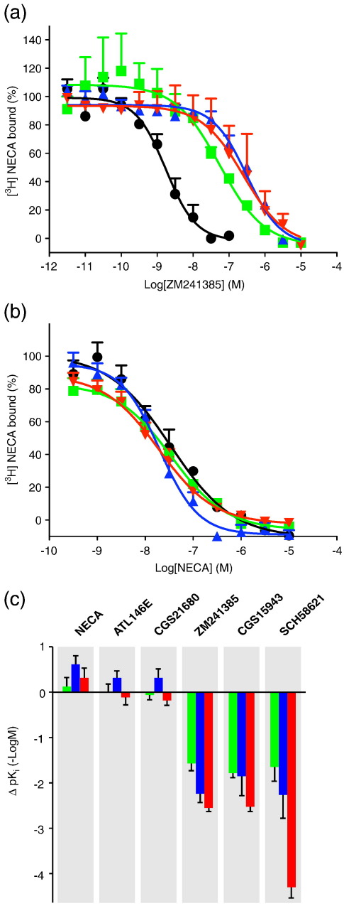
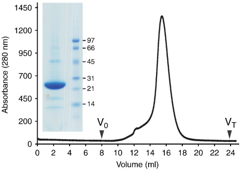
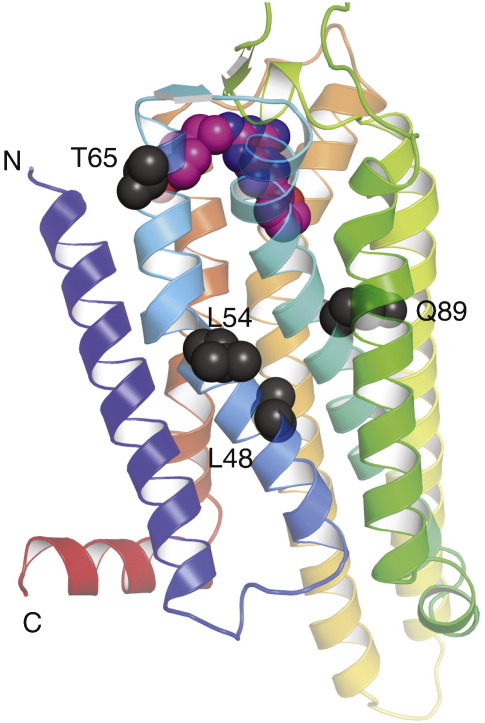
The adenosine A(2A) receptor (A(2A)R) is a G-protein-coupled receptor that plays a key role in transmembrane signalling mediated by the agonist adenosine. The structure of A(2A)R was determined recently in an antagonist-bound conformation, which was facilitated by the T4 lysozyme fusion in cytoplasmic loop 3 and the considerable stabilisation conferred on the receptor by the bound inverse agonist ZM241385. Unfortunately, the natural agonist adenosine does not sufficiently stabilise the receptor for the formation of diffraction-quality crystals. As a first step towards determining the structure of A(2A)R bound to an agonist, the receptor was thermostabilised by systematic mutagenesis in the presence of the bound agonist [(3)H]5'-N-ethylcarboxamidoadenosine (NECA). Four thermostabilising mutations were identified that when combined to give mutant A(2A)R-GL26, conferred a greater than 200-fold decrease in its rate of unfolding compared to the wild-type receptor. Pharmacological analysis suggested that A(2A)R-GL26 is stabilised in an agonist-bound conformation because antagonists bind with up to 320-fold decreased affinity. None of the thermostabilising mutations are in the ZM241385 binding pocket, suggesting that the mutations affect ligand binding by altering the conformation of the receptor rather than through direct interactions with ligands. A(2A)R-GL26 shows considerable stability in short-chain detergents, which has allowed its purification and crystallisation.


Figures

None
Graphical abstract
Fig. 1
Fig. 1
Positions of thermostabilising mutations in the primary sequence of human A2AR. The snake plot depicts the secondary structure elements found in the structure of A2AR, with the approximate position of the lipid bilayer shown in grey. (a) Thermostabilising mutations in the [3H]NECA-bound conformation are shown in orange. Mutations identified previously from [3H]NECA assays performed after heating the unliganded receptor are shown in red. Mutations that were selected by both assays are blue. (b) The 16 most thermostabilising mutations of the [3H]NECA-bound conformation of A2AR were re-assayed for thermostability in the antagonist-bound conformation using [3H]ZM241385 (Table 1): magenta, mutants that did not bind antagonist in this assay; green, mutants that are less stable than WT A2AR in the antagonist-bound conformation; and brown, mutants that are more stable than WT in the antagonist-bound conformation.
Fig. 2
Fig. 2
Thermostability of agonist-bound, DDM-solubilised A2AR and thermostabilised mutants. (a) Additive effect of thermostabilising mutations used to generate A2AR-GL23. Predicted Tm values were calculated by adding the ΔTm for each mutation (Table 1) to the apparent Tm of WT A2AR. The broken line correlates with perfect additivity. WT A2AR is represented by a black diamond. The stabilities of the best thermostable mutants containing one, two or three point mutations are labelled, respectively, as follows: L48A (GL0), green diamond; L48A-Q89A (GL10), dark-blue diamond; and L48A-Q89A-T65A (GL23), red diamond. Other double mutants (light-blue diamonds) and triple mutants (light-red diamonds) that were less stable than the optimal combinations are also shown. (b) Thermostability assays were performed on receptors partially purified in 0.025% DDM and with [3H]NECA bound; A2AR (black circles), apparent Tm of 28.6 ± 0.2 °C, n = 8; GL0 (green squares), apparent Tm of 42.2 ± 1.0 °C, n = 5; GL10 (blue triangles), apparent Tm of 46.7 ± 0.4 °C, n = 3; and GL23 (red inverted triangles), apparent Tm of 49.9 ± 0.1 °C, n = 2. (c) Stability of mutants compared to WT A2AR based on t1/2 values calculated from (b) to allow the improvement in stability of the mutants compared to WT to be calculated: GL0, 49-fold; GL10, 136-fold; and GL23, 226-fold. The colour code is the same as in (b).
Fig. 3
Fig. 3
Thermostability of the mutants GL23 and GL26. (a) Thermostability of GL26 (red squares; apparent Tm of 44.5 ± 0.8 °C, n = 3) and GL23 (blue circles; apparent Tm of 42.2 ± 0.3 °C, n = 2) after partial purification in DM (0.17%), both with [3H]NECA bound. (b) The thermostability of GL26 with [3H]NECA bound was determined by partially purifying the receptor in different detergents. GL26 was solubilised in DM and immobilised on Ni2+-NTA agarose, and then detergent exchange was performed. The results are from a single experiment performed in triplicate, with the final concentration of detergent indicated: 0.39% decanoyl-N-hydroxyethylglucamide (pink inverted triangles), apparent Tm of 42.3 °C; 0.17% decylmaltoside (red squares), apparent Tm of 42.0 °C; 0.3% NG (green inverted triangles), apparent Tm of 34.6 °C; 0.52% foscholine-10 (blue triangles), apparent Tm of 33.1 °C; 0.42% octylthioglucoside (orange circles), apparent Tm of 30.5 °C; 0.37% polyoxyethylene C8E4 (pale blue circles), apparent Tm of 26.4 °C.
Fig. 4
Fig. 4
Affinities of agonists and antagonists for A2AR and the thermostabilised mutants. (a and b) Competition binding experiments were performed by measuring the displacement of [3H]NECA bound to receptors in CHO cell membranes. Experiments were performed using three agonists (NECA, ATL146e and CGS21680) and two antagonists (CGS15943 and SCH58621) and the inverse agonist ZM241385 with example curves shown for ZM241385 (a) and NECA (b); WT A2AR, black circles; GL0, green squares; GL23, blue triangles; and GL26, red inverted triangles. Full data are shown in Table 3. (c) The differences in affinities (ΔpKi) between the WT A2AR and each of the mutants for the ligands tested were calculated from the pKi values determined in Table 3; GL0, green; GL23, blue; and GL26, red.
Fig. 5
Fig. 5
Purification of A2AR-GL31. The unglycosylated mutant of A2AR-GL26, A2AR-GL31, was expressed in insect cells using a recombinant baculovirus and purified on Ni2+-NTA. The receptor was further purified by size-exclusion chromatography (A280 trace is shown; void (V0) and total (VT) column volumes are indicated); this gave a symmetrical peak, which indicated that the preparation was monodisperse and homogenous. A Coomassie-blue-stained SDS-polyacrylamide gel (inset) showed that A2AR-GL31 represented a single band on the gel (left-hand lane) that was sufficiently pure for crystallisation (molecular weight markers are shown on the right).
Fig. 6
Fig. 6
Positions of the thermostabilising mutations in the antagonist-bound A2A structure. The structure of A2AR-StaR2 (Protein Data Bank code 3PWH) thermostabilised in an antagonist-bound conformation is shown in rainbow colouration with the N-terminus and C-terminus labelled (N and C, respectively) and the bound antagonist ZM241385 depicted as a space-filling model (C, pink: N, blue; and O, red). The four amino acid residues mutated in A2AR to generate the thermostable mutant GL26 (L48A, Q89A, T65A and A54L) are depicted as space-filling models (dark grey). Note that the amino acid sequence shown is that of A2AR-StaR2, which also contains the A54L thermostabilising mutation, whereas Leu48, Gln89 and Thr65 are identical with WT A2AR.

References

    1. Gloriam D.E., Fredriksson R., Schioth H.B. The G protein-coupled receptor subset of the rat genome. BMC Genomics. 2007;8:338. - PMC - PubMed
    1. Kobilka B.K., Deupi X. Conformational complexity of G-protein-coupled receptors. Trends Pharmacol. Sci. 2007;28:397–406. - PubMed
    1. DeWire S.M., Ahn S., Lefkowitz R.J., Shenoy S.K. Beta-arrestins and cell signaling. Annu. Rev. Physiol. 2007;69:483–510. - PubMed
    1. Hopkins A.L., Groom C.R. The druggable genome. Nat. Rev., Drug Discov. 2002;1:727–730. - PubMed
    1. Conn P.J., Christopoulos A., Lindsley C.W. Allosteric modulators of GPCRs: a novel approach for the treatment of CNS disorders. Nat. Rev., Drug Discov. 2009;8:41–54. - PMC - PubMed

Publication types

Substances