. 2011 May 15;203(10):1378-87.
doi: 10.1093/infdis/jir036.

High frequency, sustained T cell responses to PARV4 suggest viral persistence in vivo

Affiliations

High frequency, sustained T cell responses to PARV4 suggest viral persistence in vivo

Ruth Simmons et al. J Infect Dis. .

Abstract

Background: Parvovirus 4 (PARV4) is a recently identified human virus that has been found in livers of patients infected with hepatitis C virus (HCV) and in bone marrow of individuals infected with human immunodeficiency virus (HIV). T cells are important in controlling viruses but may also contribute to disease pathogenesis. The interaction of PARV4 with the cellular immune system has not been described. Consequently, we investigated whether T cell responses to PARV4 could be detected in individuals exposed to blood-borne viruses.

Methods: Interferon γ (IFN-γ) enzyme-linked immunospot assay, intracellular cytokine staining, and a tetrameric HLA-A*0201-peptide complex were used to define the lymphocyte populations responding to PARV4 NS peptides in 88 HCV-positive and 13 HIV-positive individuals. Antibody responses were tested using a recently developed PARV4 enzyme-linked immunosorbent assay.

Results: High-frequency T cell responses against multiple PARV4 NS peptides and antibodies were observed in 26% of individuals. Typical responses to the NS pools were >1000 spot-forming units per million peripheral blood mononuclear cells.

Conclusions: PARV4 infection is common in individuals exposed to blood-borne viruses and elicits strong T cell responses, a feature typically associated with persistent, contained infections such as cytomegalovirus. Persistence of PARV4 viral antigen in tissue in HCV-positive and HIV-positive individuals and/or the associated activated antiviral T cell response may contribute to disease pathogenesis.


Figures
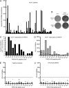
Figure 1.
Figure 1.
Analysis of PARV4 T cell responses in HCV-infected patients and controls. T cell responses were assessed by IFN-γ ELISpot assays with PARV4 NS peptides. (A) T cells from 88 HCV-infected patients to each peptide pool are summed so that each column represents the response of one patient to the entire NS protein. (B) These photos of ELISpot wells are examples of the highest levels of responses and their negative control wells (DMSO). Peptide pool responses for patients 117 and 425 are 410 and 248 SFU/well, respectively. (CD) These graphs show the distribution and the magnitude of the T cell responses across the NS protein. (C) Each column represents the number of patients responding to each pool. These are concentrated at the N-terminus of the protein. (D) Each column represents the range of responses of all responders and the median to that peptide pool. PARV4 NS responders indicate preferential recognition of N-terminal pools of peptides. (EF) T cell responses of 9 healthy individuals (E) and 7 B19-positive patients (F) to PARV4 NS peptide pools are displayed. Each point represents the T cell response of one individual to the peptide pool on the x-axis. The highest response was 35 SFU/106 PBMC. The dashed line marks the cutoff for positivity.
Figure 2.
Figure 2.
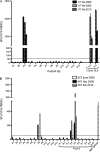
Longitudinal analysis of T cell responses to PARV4 NS. T cell responses of patients 117 (A) and 457 (B) are illustrated here as examples of sustained responses over time. The dates next to the patient numbers represent the sample date. (”457 june 2008” sample: these cells were cultured with peptide for 22 days before the assay.) OLP, overlapping peptides.
Figure 3.
Figure 3.
T cell responses to PARV4 NS protein in HIV-infected individuals. Results are displayed as in Figures 1E–1F. Three of 13 HIV-positive patients had a positive response to PARV4 NS peptide pools (N067, N090, and R010, with CD4+ T cell counts of 430, 350, and 460 cells/μL, respectively), and the distribution of responses across the NS protein is shown. Individuals with no responses had CD4+ T cell counts between 130 and 1300 cells/μL (median: 430 cells/μL).
Figure 4.
Figure 4.
Demonstration of PARV4 NS peptides triggering the production of IFN-γ by CD8+ T cells. Short-term PBMC lines for patients 314 (upper panel) and 457 (lower panel) elicit a CD8+ T cell response to 2 pools of peptides and one 9mer epitope, respectively. The control peptide is a tetanus toxin peptide [31].
Figure 5.
Figure 5.
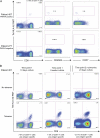
HLA-A*0201–restricted PARV4 RMTENIVEV-specific T cells have an effector memory phenotype and proliferate in culture in vitro. (A) FACS plots for patient 457 (top panel), from whom the epitope was determined (HLA-A*0201 and positive response for peptides 8.4–8.5), and for patient 277 (lower panel, ”negative” = no response on ELISpot assays to PARV4 NS). Left-hand plots are gated on CD3+ cells. The middle and right-hand panels assess the phenotype of the epitope-specific cells and are gated on CD3+ CD8+ T cells. CD45RA and CCR7 are used as memory markers. Antigen-specific cells are visible in patient 457 at a frequency of .19% and do not present CD45RA or CCR7 on their surface. (B) After short-term culture, tetramer HLA-A*02-RMT–positive cells have expanded to 7% of CD8+ T cells. This appears to plateau as the level is unchanged after 6 weeks (middle panels). Tetramer-positive cells are still visible at a second time point (right panels) in the same patient, although the percentage decreases. Samples from time points 1 and 2 were taken 20 months apart.

Similar articles

Cited by

References

    1. Brown KE. The expanding range of parvoviruses which infect humans. Rev Med Virol. 2010;20:231–44. - PubMed
    1. Jones MS, Kapoor A, Lukashov VV, Simmonds P, Hecht F, Delwart E. New DNA viruses identified in patients with acute viral infection syndrome. J Virol. 2005;79:8230–6. - PMC - PubMed
    1. Schneider B, Fryer JF, Reber U, et al. Persistence of novel human parvovirus PARV4 in liver tissue of adults. J Med Virol. 2008;80:345–51. - PubMed
    1. Manning A, Willey SJ, Bell JE, Simmonds P. Comparison of tissue distribution, persistence, and molecular epidemiology of parvovirus B19 and novel human parvoviruses PARV4 and human bocavirus. J Infect Dis. 2007;195:1345–52. - PMC - PubMed
    1. Longhi E, Bestetti G, Acquaviva V, et al. Human parvovirus 4 in the bone marrow of Italian patients with AIDS. AIDS. 2007;21:1481–3. - PubMed

Publication types

MeSH terms