

Structure and function of cytochromes P450 2B: from mechanism-based inactivators to X-ray crystal structures and back
- PMID: 21502194
- PMCID: PMC3127236
- DOI: 10.1124/dmd.111.039719
Structure and function of cytochromes P450 2B: from mechanism-based inactivators to X-ray crystal structures and back
Abstract
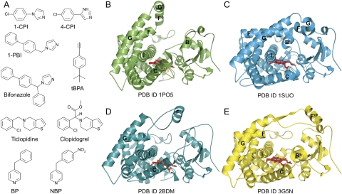
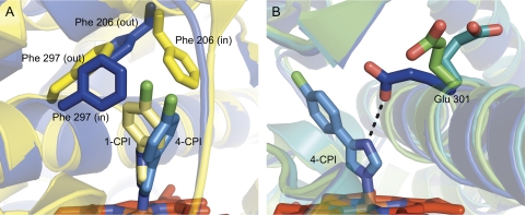
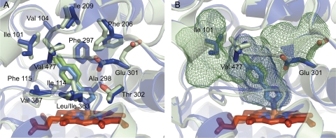
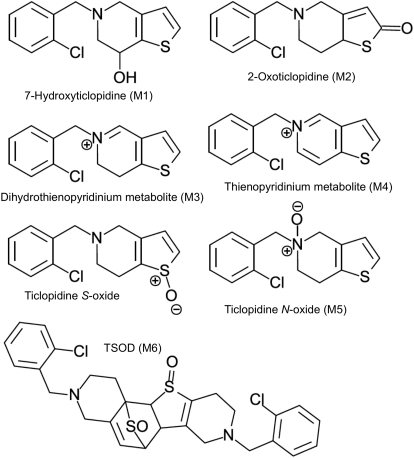
This article reviews work from the author dating back to 1978 and focuses on the structural basis of cytochrome P450 (P450) function using available contemporary techniques. Early studies used mechanism-based inactivators that bound to the protein moiety of hepatic P450s to try to localize the active site. Subsequent studies used cDNA cloning, heterologous expression, site-directed mutagenesis, and homology modeling based on multiple bacterial P450 X-ray crystal structures to predict the active sites of CYP2B enzymes with considerable accuracy. Breakthroughs in engineering and expression of mammalian P450s enabled us to determine our first X-ray crystal structure of ligand-free rabbit CYP2B4. To date, we have solved 11 CYP2B4 and three human CYP2B6 structures, which represent four significantly different conformations. The plasticity of CYP2B4 has been confirmed by deuterium exchange mass spectrometry and is substantiated by molecular dynamics simulations. In addition to major movement of secondary structure elements, more subtle reorientation of active site side chains, especially Phe206, Phe297, and Glu301, contributes to the ability of CYP2B enzymes to bind various ligands. Isothermal titration calorimetry has proven to be a useful tool for studying the thermodynamics of ligand binding to CYP2B4 and CYP2B6, and NMR has enabled study of ligand binding orientation in solution as an adjunct to X-ray crystallography. A major challenge remains to harness the power of the various approaches to facilitate prediction of CYP2B specificity and inhibition.
Figures
References
-
- Blobaum AL, Harris DL, Hollenberg PF. (2005) P450 active site architecture and reversibility: inactivation of cytochromes P450 2B4 and 2B4 T302A by tert-butyl acetylenes. Biochemistry 44:3831–3844 - PubMed
-
- Cameron MD, Wen B, Allen KE, Roberts AG, Schuman JT, Campbell AP, Kunze KL, Nelson SD. (2005) Cooperative binding of midazolam with testosterone and alpha-naphthoflavone within the CYP3A4 active site: a NMR T1 paramagnetic relaxation study. Biochemistry 44:14143–14151 - PubMed
-
- Cameron MD, Wen B, Roberts AG, Atkins WM, Campbell AP, Nelson SD. (2007) Cooperative binding of acetaminophen and caffeine within the P450 3A4 active site. Chem Res Toxicol 20:1434–1441 - PubMed
-
- Ciaccio PJ, Duignan DB, Halpert JR. (1987) Selective inactivation by chloramphenicol of the major phenobarbital-inducible isozyme of dog liver cytochrome P-450. Drug Metab Dispos 15:852–856 - PubMed
-
- Cosme J, Johnson EF. (2000) Engineering microsomal cytochrome P450 2C5 to be a soluble, monomeric enzyme. Mutations that alter aggregation, phospholipid dependence of catalysis, and membrane binding. J Biol Chem 275:2545–2553 - PubMed
Publication types
MeSH terms
Substances
Grants and funding
LinkOut - more resources
Full Text Sources
