Review
. 2011 Apr 18;193(2):257-66.
doi: 10.1083/jcb.201010131.

Manifestations and mechanisms of stem cell aging

Affiliations
Review

Manifestations and mechanisms of stem cell aging

Ling Liu et al. J Cell Biol. .

Abstract

Adult stem cells exist in most mammalian organs and tissues and are indispensable for normal tissue homeostasis and repair. In most tissues, there is an age-related decline in stem cell functionality but not a depletion of stem cells. Such functional changes reflect deleterious effects of age on the genome, epigenome, and proteome, some of which arise cell autonomously and others of which are imposed by an age-related change in the local milieu or systemic environment. Notably, some of the changes, particularly epigenomic and proteomic, are potentially reversible, and both environmental and genetic interventions can result in the rejuvenation of aged stem cells. Such findings have profound implications for the stem cell-based therapy of age-related diseases.


Figures

Figure 1.
Figure 1.
The chronological and replicative lifespan of stem cells. During prolonged periods of quiescence and by the process of self-renewal to establish a cellular continuum, stem cells experience chronological aging caused by the accumulation of damaged or aberrant intracellular molecules. During the process of asymmetric cell division and self-renewal, stem cells also experience replicative aging, which is particularly important in tissues with high turnover rates.
Figure 2.
Figure 2.
Decline in stem cell function with age. In young animals, stem cells divide asymmetrically to self-renew and give rise to lineage-specific differentiated progeny during tissue homeostasis or regeneration. With age, some stem cells lose their lineage specificity and give rise to nonfunctional progeny, resulting in loss of tissue integrity and decline of physiological function, even though the number of stem cells remains unaffected. Some stem cells lose the capacity for self-renewal, resulting in symmetric cell divisions giving rise to two differentiated daughters and a gradual depletion of the stem cell pool. The senescence of stem cells can also contribute to a loss of functional stem cells. The increase in malignancies with age, particularly in epithelia with high turnover rates, has been proposed to arise from within the stem cell compartment or from early progenitors.
Figure 3.
Figure 3.
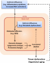
Extrinsic and intrinsic influences on stem cell aging. Age-related changes in the systemic milieu, for example, an increased level of inflammatory cytokines or Wnt pathway activators in the circulation, can lead to changes of signaling cascades and molecular changes within cells. For stem cells, the effects of extrinsic influences may also be transmitted indirectly via detrimental changes in the niche. Meanwhile, intrinsic changes, such as changes in mitochondrial activity or metabolic rate, can also occur during cellular aging. The extrinsic and intrinsic influences converge at intracellular molecular effectors, such as reactive oxygen species (ROS), which can cause either reversible changes (such as protein oxidation) or irreversible changes (such as DNA mutations) to macromolecules in the cell. The combinatorial effects of genomic, epigenomic, and proteomic changes lead to a decline in cellular function, which in turn contributes to tissue dysfunction and organismal aging. Because dysfunctional stem cells give rise to abnormal differentiated cells in the tissue, stem cell aging exacerbates the extrinsic influences of aging, thereby also contributing to the aging process of the tissue and organism.

References

    1. Akbari M., Krokan H.E. 2008. Cytotoxicity and mutagenicity of endogenous DNA base lesions as potential cause of human aging. Mech. Ageing Dev. 129:353–365 10.1016/j.mad.2008.01.007 - DOI - PubMed
    1. Andl T., Reddy S.T., Gaddapara T., Millar S.E. 2002. WNT signals are required for the initiation of hair follicle development. Dev. Cell. 2:643–653 10.1016/S1534-5807(02)00167-3 - DOI - PubMed
    1. Barker N., van Es J.H., Kuipers J., Kujala P., van den Born M., Cozijnsen M., Haegebarth A., Korving J., Begthel H., Peters P.J., Clevers H. 2007. Identification of stem cells in small intestine and colon by marker gene Lgr5. Nature. 449:1003–1007 10.1038/nature06196 - DOI - PubMed
    1. Beerman I., Bhattacharya D., Zandi S., Sigvardsson M., Weissman I.L., Bryder D., Rossi D.J. 2010. Functionally distinct hematopoietic stem cells modulate hematopoietic lineage potential during aging by a mechanism of clonal expansion. Proc. Natl. Acad. Sci. USA. 107:5465–5470 10.1073/pnas.1000834107 - DOI - PMC - PubMed
    1. Bell D.R., Van Zant G. 2004. Stem cells, aging, and cancer: inevitabilities and outcomes. Oncogene. 23:7290–7296 10.1038/sj.onc.1207949 - DOI - PubMed

Publication types