

Editorial
doi: 10.1148/radiol.11101800.
CT dose index and patient dose: they are not the same thing
- PMID: 21502387
- PMCID: PMC3079120
- DOI: 10.1148/radiol.11101800
Item in Clipboard
Editorial
CT dose index and patient dose: they are not the same thing
Radiology.
2011 May.
Abstract
Estimates of individual patient risk, and epidemiologic studies assessing potential late effects, must use patient size–specific dose estimates—they cannot use only scanner output (volume CT dose index or dose-length product).
Figures
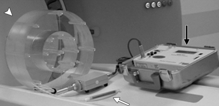
Equipment typically used to measure CTDI100 includes an integrating electrometer (black arrow), a 100-mm-long CTDI ionization chamber (white arrow), and a CTDI phantom made of polymethylmethacrylate (arrowhead). The phantom is placed with its long axis perpendicular to the plane of the transverse CT scan and the ion chamber placed in one of the holes through the phantom. CTDI100 is obtained by integrating the dose over 100 mm from a single transverse scan and dividing it by the nominal beam width. (Reprinted, with permission, from reference .)
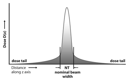
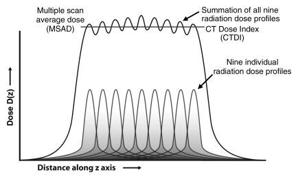
(a) Radiation dose profile along a line perpendicular to the scan plane shows a peak dose level at the center of the primary beam and long dose tails caused by scattered radiation. NT = nominal beam width. (b) The radiation dose profiles from nine adjacent transverse CT scans along a line perpendicular to the transverse scans, when summed, produce the MSAD profile. The value of MSAD is the average value of this profile over one scan interval in the central portion of the profile. (Reprinted, with permission, from reference .)
(a) Radiation dose profile along a line perpendicular to the scan plane shows a peak dose level at the center of the primary beam and long dose tails caused by scattered radiation. NT = nominal beam width. (b) The radiation dose profiles from nine adjacent transverse CT scans along a line perpendicular to the transverse scans, when summed, produce the MSAD profile. The value of MSAD is the average value of this profile over one scan interval in the central portion of the profile. (Reprinted, with permission, from reference .)
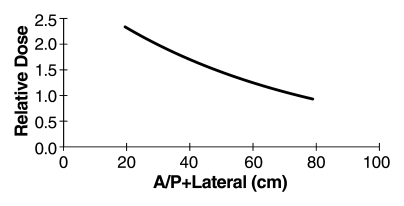
Graph shows relative dose (mean patient dose per 1 mGy of scanner output, CTDIvol) for an abdominal CT scan and different patient sizes (here represented by the sum of anteroposterior [A/P] and lateral dimensions). Over the range of patient sizes from a newborn to a large adult, relative dose is exponentially related to patient size. For a patient with an anteroposterior dimension of 30 cm and a lateral dimension of 40 cm, the anteroposterior + lateral value would be 70 cm and the mean patient dose in the center of the scan range would be approximately equivalent to the CTDIvol value reported on the console. For a neonate having both anteroposterior and lateral dimensions of 10 cm, the anteroposterior + lateral value would be 20 cm and the mean patient dose in the center of an abdomen scan would be about 2.3 times the displayed CTDI value, for body CTDIvol measurements made by using a 32-cm phantom. CTDIvol measurements made on the basis of 16-cm phantoms would require a different scale factor.
Comment in
-
CTDIvol, DLP, and effective dose are excellent measures for use in CT quality improvement.Radiology. 2011 Dec;261(3):999; author reply 999-1000. doi: 10.1148/radiol.11111055. Radiology. 2011. PMID: 22096003 Free PMC article. No abstract available.
References
-
- Shope TB, Gagne RM, Johnson GC. A method for describing the doses delivered by transmission x-ray computed tomography. Med Phys 1981;8(4):488–495 - PubMed
-
- American Association of Physicists in Medicine Standardized methods for measuring diagnostic x-ray exposures. New York, NY: American Association of Physicists in Medicine, 1990
-
- American Association of Physicists in Medicine The measurement, reporting and management of radiation dose in CT. Report 96. AAPM Task Group 23 of the Diagnostic Imaging Council CT Committee. College Park, Md: American Association of Physicists in Medicine, 2008
-
- European Commission European guidelines on quality criteria for computed tomography. EUR 16262 EN. Luxembourg: European Commission, 2000
-
- International Electrotechnical Commission Medical electrical equipment. Part 2-44: Particular requirements for the safety of x-ray equipment for computed tomography. IEC publication no. 60601-2-44 Ed. 2.1 Geneva, Switzerland: International Electrotechnical Commission Central Office, 2002
Publication types
MeSH terms
Grants and funding
LinkOut - more resources
Full Text Sources
Medical
