. 2011 May 3;108(18):7517-22.
doi: 10.1073/pnas.1016428108. Epub 2011 Apr 18.

Three chemokine receptors cooperatively regulate homing of hematopoietic progenitors to the embryonic mouse thymus

Affiliations

Three chemokine receptors cooperatively regulate homing of hematopoietic progenitors to the embryonic mouse thymus

Lesly Calderón et al. Proc Natl Acad Sci U S A. .

Abstract

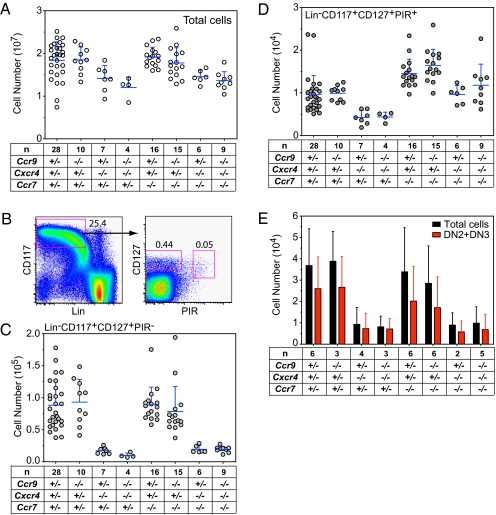
The thymus lacks self-renewing hematopoietic cells, and thymopoiesis fails rapidly when the migration of progenitor cells to the thymus ceases. Hence, the process of thymus homing is an essential step for T-cell development and cellular immunity. Despite decades of research, the molecular details of thymus homing have not been elucidated fully. Here, we show that chemotaxis is the key mechanism regulating thymus homing in the mouse embryo. We determined the number of early thymic progenitors in the thymic rudiments of mice deficient for one, two, or three of the chemokine receptor genes, chemokine (C-C motif) receptor 9 (Ccr9), chemokine (C-C motif) receptor 7 (Ccr7), and chemokine (C-X-C motif) receptor 4 (Cxcr4). In the absence of all three chemokine receptors, thymus homing was reduced about 100-fold both before and after vascularization of the thymic rudiment. In the absence of only two of these three chemokine receptor genes, thymus homing was much less affected (only two- to 10-fold), indicating that the chemotactic regulation of thymus homing is remarkably robust. Our results reveal the redundant roles of Ccr9, Ccr7, and Cxcr4 for thymic homing and provide a framework to examine the regulation of progenitor homing in the postnatal thymus.


Conflict of interest statement

The authors declare no conflict of interest.

Figures

Fig. 1.
Fig. 1.
Combined lack of Ccr9 and Cxcr4 chemokine receptors does not abolish intrathymic T-cell development. The number of thymocytes is shown for embryos of the indicated genotypes at E14.5 and E17.5. The number of analyzed embryos is indicated (n).
Fig. 2.
Fig. 2.
Spatial distribution of CD45+ cells in the pharyngeal region of E12.5 embryos. Cryosections were stained for cytokeratin 8 (green fluorescence) and CD45 (red fluorescence). The boundaries between the anlagen of the parathyroid (pointing to the upper left corner) and the thymus (lower right) are indicated by the dashed lines. The genotypes of embryos are indicated above AH. The panels are representative of two to four thymic lobes. (Scale bar, 50 μm.)
Fig. 3.
Fig. 3.
T-cell progenitors in the thymus of E14.5 embryos deficient for chemokine receptors. (A) Total number of thymocytes in embryos of the indicated genotypes. (B) Representative flow cytometric profile of a Ccr9+/−;Ccr7+/−;Cxcr4+/− embryo to illustrate the identification of ETPs. Numbers represent percentages of total thymocytes. (C) Number of ETPs in embryos of the indicated genotypes. Note that the scale is expanded in the lower part of the y axis; the inset is a magnification to show the absolute number of ETPs for the three color-coded genotypes. Average values are indicated; each data point represents one embryo, and the total number of embryos per genotype is indicated (n).
Fig. 4.
Fig. 4.
Early progenitor cells in the fetal liver of E14.5 embryos. (A) Total number of cells for embryos of the indicated genotypes. (B) Representative flow cytometric profile of a Ccr9+/−;Ccr7+/−;Cxcr4+/− embryo to illustrate the identification of LinCD117+CD127+PIR and LinCD117+CD127+PIR+ populations. Numbers refer to percentages of total cells. (C) Number of LinCD117+CD127+PIR cells in the fetal liver of embryos with the indicated genotypes. (D) Number of LinCD117+CD127+PIR+ cells. (E) Total number of cells and number of DN2 + DN3 cells obtained after in vitro culture of 100 LinCD117+CD127+PIR+ cells for 8 d on OP9-DL1 stroma. The total number of embryos analyzed per genotype is indicated (n).
Fig. 5.
Fig. 5.
T-cell progenitors in the thymus of E17.5 embryos deficient for chemokine receptors. (AE) Distribution of CD45+ cells in the thymic rudiment of E17.5 embryos. The genotype is indicated at the top of each panel. Cryosections were stained for cytokeratin 8 (epithelial marker, green fluorescence), CD45 (marker of hematopoietic cells, red fluorescence), and CD31 (endothelial marker, blue fluorescence). The panels are representative of two to six thymic lobes. (Scale bar, 100 μm.) (F) Number of total thymocytes in embryos of the indicated genotypes. (G) Representative flow cytometric profile of a Ccr9+/−;Ccr7+/−;Cxcr4+/− embryo to illustrate the identification ETPs. Numbers refer to percentages of total thymocytes. (H) Number of ETPs in embryos of the indicated genotypes. Note that the scale is expanded in the lower part of the y axis. The total number of embryos per genotype is indicated (n).

References

    1. Owen JJ, Ritter MA. Tissue interaction in the development of thymus lymphocytes. J Exp Med. 1969;129:431–442. - PMC - PubMed
    1. Moore MA, Owen JJ. Experimental studies on the development of the thymus. J Exp Med. 1967;126:715–726. - PMC - PubMed
    1. Ritter MA. Embryonic mouse thymus development: Stem cell entry and differentiation. Immunology. 1978;34:69–75. - PMC - PubMed
    1. Pyke KW, Bach JF. The in vitro migration of murine fetal liver cells to thymic rudiments. Eur J Immunol. 1979;9:317–323. - PubMed
    1. Jotereau FV, Houssaint E, Le Douarin NM. Lymphoid stem cell homing to the early thymic primordium of the avian embryo. Eur J Immunol. 1980;10:620–627. - PubMed

Publication types

Substances