. 2011 May 3;108(18):7391-6.
doi: 10.1073/pnas.1018279108. Epub 2011 Apr 18.

Conserved, developmentally regulated mechanism couples chromosomal looping and heterochromatin barrier activity at the homeobox gene A locus

Affiliations

Conserved, developmentally regulated mechanism couples chromosomal looping and heterochromatin barrier activity at the homeobox gene A locus

Yoon Jung Kim et al. Proc Natl Acad Sci U S A. .

Erratum in

  • Proc Natl Acad Sci U S A. 2011 Nov;108(47):19096

Abstract

Establishment and segregation of distinct chromatin domains are essential for proper genome function. The insulator protein CCCTC-binding factor (CTCF) is involved in creating boundaries that segregate chromatin and functional domains and in organizing higher-order chromatin structures by promoting chromosomal loops across the vertebrate genome. Here, we investigate the insulation properties of CTCF at the human and mouse homeobox gene A (HOXA) loci. Although cohesin loading at the CTCF binding site is required for looping, we found that cohesin is dispensable for chromatin barrier activity at that site. Using mouse embryonic stem cells in both a pluripotent and differentiated neuronal progenitor state, we determined that embryonic stem cell pluripotency factor OCT4 antagonizes cohesin loading at the CTCF binding site. Loss of OCT4 in the committed and differentiated neuronal progenitor cells results in loading of cohesin and chromosome looping, which contributes to heterochromatin partitioning and selective gene activation across the HOXA locus. Our analysis reveals that chromatin barrier activity of CTCF is evolutionarily conserved and is responsible for the coordinated establishment of chromatin structure, higher-order architecture, and developmental expression of the HOXA locus.


Conflict of interest statement

The authors declare no conflict of interest.

Figures
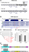
Fig. 1.
Fig. 1.
CBS5 functions as a chromatin barrier in the HOXA locus. A represents the HOXA gene expression status and CTCF binding sites in ES and differentiated cells. B is the result of CBS5 sequence alignment; the red box is the core 20 bp of CBS5 and the blue box is a candidate site for OCT4 binding in mouse HOXA locus. C is a phylogenetic tree indicating conservation of the CBS5 sequence primarily in mammals. D is the percentage of EGFP expression levels normalized to the gamma actin (ACTG1) gene expression, determined by RT-qPCR, upon expression of LacI or LacI-EMD proteins, for cells containing either a control reporter or a reporter containing the CBS5 element between LacO and the EGFP gene. Schematics of the CBS5 and control reporters are shown in E.
Fig. 2.
Fig. 2.
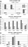
CTCF function in chromatin barrier activity and looping. CTCF knockdown was performed in IMR90 cells to investigate the function of CTCF in chromatin barrier activity and looping. A shows qRT-PCR results of CTCF and RAD21 (cohesin subunit) ChIP analysis of chromatin isolated from IMR90 cells infected with either a control virus, pGIPz (IMR90), or a CTCF knockdown virus (CTCF KD). Dark-gray bars represent enrichment level of target protein at CBS5, whereas light-gray bars represent the enrichment at a random control site. Inset is Western blot analysis showing the efficacy of CTCF KD in IMR90 cells. B is the ChIP-qPCR results using H3K27me3 antibody, and shows the H3K27me3 levels at CTCF binding sites (CBS4 to CBS7) including CBS5 in the HOXA locus. C shows the relative expression level of HOXA genes near CBS5 (two upstream genes, HOXA6 and HOXA7, and downstream genes, HOXA9 and HOXA10), in IMR90 cells infected with CTCF KD and control (pGIPz) virus, respectively. The y axes represent the cDNA copy number of HOXA genes normalized by the cDNA copy number of the control ACTG1 gene (HOXA copy no./ACTG1 copy no.  × 108).
Fig. 3.
Fig. 3.
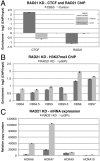
Cohesin function in chromatin barrier activity and looping. RAD21 knockdown was performed in IMR90 to determine the role of cohesin in chromatin barrier activity and looping independent of CTCF. A and B are ChIP-qPCR results using CTCF, RAD21, and H3K27me3 antibody in IMR90 cells infected with RAD21 knockdown virus (RAD21 KD). Inset is Western blot analysis showing the efficacy of RAD21 KD in IMR90 cells. In B, the H3K27me3 levels at CTCF binding sites (CBS4 to CBS7) in the HOXA locus were determined by ChIP-qPCR. C shows the mRNA expression level (relative copy number) of HOXA genes (HOXA6HOXA13) in control (pGIPz) and RAD21 KD cells. The y axes represent the cDNA copy number of HOXA genes normalized by the cDNA copy number of the control gamma actin gene, ACTG1 (HOXA copy no./ACTG1 copy no.  × 108).
Fig. 4.
Fig. 4.
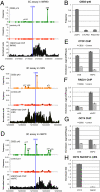
Chromosome architecture of the human and mouse HOXA locus. Interaction frequencies between the anchor primer (noted by a yellow circle) and other primers in the locus were determined by real-time PCR and normalized to a random ligation library generated from EcoRI restriction digestion fragments from the bacterial artificial clones (BAC) covering the HOXA locus (CTD-3054H22 and CTD-2536K9) of this locus. A shows the interaction frequencies observed with the anchor primer located at CBS5 (blue vertical bar) with the all the other primers in the locus in the top panel. The middle panel shows the interaction frequencies observed with a control anchor primer located 12-Kb downstream from CBS5 with all the other primers in the locus. The bottom panel shows the corresponding H3K27me3 ChIP-seq density across the HOXA locus in IMR90 cells. B is the result of the 3C assay in IMR90 cells infected with either CTCF or RAD21 knockdown (KD) viruses, or control pGIPz virus, tested to determine the interaction between CBS5 and the heterochromatin boundary (p40), which was detected as the strongest specific long-range interaction site in A. C and D show the interaction frequency of CBS5 or control primer (noted by yellow circles) with all the other 3C primers in mES cells and mNPC across the mouse HOXA locus. The interaction frequencies were calculated as in A. The control random ligation library for the mouse HOXA locus was generated using the BAC, RP23-33N14. The H3K27me3 ChIP-Seq densities across the HOXA locus from mES and mNPC are shown in the bottom panel. A strong interaction frequency observed between the close proximal site (within 10-Kb downstream to CBS5) and the anchor site at CBS5 likely results from self-ligation and thus the strong signal may not indicate long-range interaction between the two sites. EG are the results of ChIP-qPCR at CBS5 performed using CTCF, RAD21, and OCT4 antibodies, respectively, and using mES and mNPC chromatin. H shows the results of ReChIP performed with CTCF and RAD21 antibodies following a primary OCT4 ChIP in ES cells.
Fig. 5.
Fig. 5.
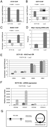
Effect of OCT4 overexpression on chromatin structure and architecture. AC are the results of ChIP-qPCR analysis of CTCF, OCT4, and RAD21, respectively, at the CBS5 site using chromatin isolated from IMR90 cells overexpressing OCT4 (OCT4 OE) or a control protein (LacZ). Western blot analysis showing the efficacy of OCT4 OE in IMR90 cells is displayed in the inset of B. D shows the interaction frequency for major looping between CBS5 and the heterochromatin boundary site (p40) in IMR90 cells overexpressing OCT4 and LacZ. In E, H3K27me3 ChIP-qPCR analysis of chromatin isolated from IMR90 cells overexpressing OCT4 (OCT4 OE) was carried out at four CTCF binding sites, CBS4, 5, 6, and 7 in the HOXA locus. F is the mRNA expression analysis on the HOXA genes (HOXA6, 7, 9, and 10) after OCT4 overexpression in IMR90 cells. Values of the y axis represent the relative copy number of HOXA cDNAs normalized to ACTG1 cDNA. G is the proposed model of regulated chromatin looping at HOXA locus.

References

    1. Phillips JE, Corces VG. CTCF: Master weaver of the genome. Cell. 2009;137:1194–1211. - PMC - PubMed
    1. Cuddapah S, et al. Global analysis of the insulator binding protein CTCF in chromatin barrier regions reveals demarcation of active and repressive domains. Genome Res. 2009;19:24–32. - PMC - PubMed
    1. Simon JA, Kingston RE. Mechanisms of polycomb gene silencing: Knowns and unknowns. Nat Rev Mol Cell Biol. 2009;10:697–708. - PubMed
    1. Defossez PA, et al. The human enhancer blocker CTC-binding factor interacts with the transcription factor Kaiso. J Biol Chem. 2005;280:43017–43023. - PubMed
    1. Docquier F, et al. Decreased poly(ADP-ribosyl)ation of CTCF, a transcription factor, is associated with breast cancer phenotype and cell proliferation. Clin Cancer Res. 2009;15:5762–5771. - PubMed

Publication types

LinkOut - more resources