

Topographical organization of the pedunculopontine nucleus
- PMID: 21503154
- PMCID: PMC3074429
- DOI: 10.3389/fnana.2011.00022
Topographical organization of the pedunculopontine nucleus
Abstract
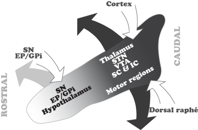
Neurons in the pedunculopontine nucleus (PPN) exhibit a wide heterogeneity in terms of their neurochemical nature, their discharge properties, and their connectivity. Such characteristics are reflected in their functional properties and the behaviors in which they are involved, ranging from motor to cognitive functions, and the regulation of brain states. A clue to understand this functional versatility arises from the internal organization of the PPN. Thus, two main areas of the PPN have been described, the rostral and the caudal, which display remarkable differences in terms of the distribution of neurons with similar phenotype and the projections that originate from them. Here we review these differences with the premise that in order to understand the function of the PPN it is necessary to understand its intricate connectivity. We support the case that the PPN should not be considered as a homogeneous structure and conclude that the differences between rostral and caudal PPN, along with their intrinsic connectivity, may underlie the basis of its complexity.
Keywords: basal ganglia; brainstem; connectivity; microcircuits; neuronal heterogeneity; pedunculopontine; reticular activating system; synaptic organization.
Figures
Similar articles
-
The Cellular Diversity of the Pedunculopontine Nucleus: Relevance to Behavior in Health and Aspects of Parkinson's Disease.Neuroscientist. 2017 Aug;23(4):415-431. doi: 10.1177/1073858416682471. Epub 2016 Dec 7. Neuroscientist. 2017. PMID: 27932591 Review.
-
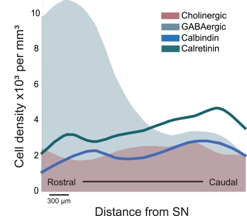
GABAergic neuron distribution in the pedunculopontine nucleus defines functional subterritories.J Comp Neurol. 2009 Aug 1;515(4):397-408. doi: 10.1002/cne.22065. J Comp Neurol. 2009. PMID: 19459217
-
Divergent motor projections from the pedunculopontine nucleus are differentially regulated in Parkinsonism.Brain Struct Funct. 2014 Jul;219(4):1451-62. doi: 10.1007/s00429-013-0579-6. Epub 2013 May 26. Brain Struct Funct. 2014. PMID: 23708060 Free PMC article.
-
Pedunculopontine nucleus: functional organization and clinical implications.Neurology. 2013 Mar 19;80(12):1148-55. doi: 10.1212/WNL.0b013e3182886a76. Neurology. 2013. PMID: 23509047 Review.
-
Glutamatergic Circuits in the Pedunculopontine Nucleus Modulate Multiple Motor Functions.Neurosci Bull. 2024 Nov;40(11):1713-1731. doi: 10.1007/s12264-024-01314-y. Epub 2024 Nov 11. Neurosci Bull. 2024. PMID: 39527367 Free PMC article.
Cited by
-
Neural mechanisms and potential treatment of epilepsy and its complications.Am J Transl Res. 2014 Nov 22;6(6):625-30. eCollection 2014. Am J Transl Res. 2014. PMID: 25628775 Free PMC article. Review.
-
On the Role of the Pedunculopontine Nucleus and Mesencephalic Reticular Formation in Locomotion in Nonhuman Primates.J Neurosci. 2016 May 4;36(18):4917-29. doi: 10.1523/JNEUROSCI.2514-15.2016. J Neurosci. 2016. PMID: 27147647 Free PMC article.
-
Imaging the effect of the circadian light-dark cycle on the glymphatic system in awake rats.Proc Natl Acad Sci U S A. 2020 Jan 7;117(1):668-676. doi: 10.1073/pnas.1914017117. Epub 2019 Dec 17. Proc Natl Acad Sci U S A. 2020. PMID: 31848247 Free PMC article.
-
The Mesoscopic Connectome of the Cholinergic Pontomesencephalic Tegmentum.Front Neuroanat. 2022 May 17;16:843303. doi: 10.3389/fnana.2022.843303. eCollection 2022. Front Neuroanat. 2022. PMID: 35655583 Free PMC article.
-
Human wildtype tau expression in cholinergic pedunculopontine tegmental neurons is sufficient to produce PSP-like behavioural deficits and neuropathology.Eur J Neurosci. 2021 Nov;54(10):7688-7709. doi: 10.1111/ejn.15496. Epub 2021 Nov 2. Eur J Neurosci. 2021. PMID: 34668254 Free PMC article.
References
-
- Alderson H. L., Latimer M. P., Winn P. (2006). Intravenous self-administration of nicotine is altered by lesions of the posterior, but not anterior, pedunculopontine tegmental nucleus. Eur. J. Neurosci. 23, 2169–2175 - PubMed
-
- Andero R., Torras-Garcia M., Quiroz-Padilla M. F., Costa-Miserachs D., Coll-Andreu M. (2007). Electrical stimulation of the pedunculopontine tegmental nucleus in freely moving awake rats: time- and site-specific effects on two-way active avoidance conditioning. Neurobiol. Learn. Mem. 87, 510–521 - PubMed
-
- Benabid A. L. (2003). Deep brain stimulation for Parkinson's disease. Curr. Opin. Neurobiol. 13, 696–706 - PubMed
Grants and funding
LinkOut - more resources
Full Text Sources