

Image quality assessment of the right ventricle with three different delayed enhancement sequences in patients suspected of ARVC/D
- PMID: 21503703
- PMCID: PMC3326369
- DOI: 10.1007/s10554-011-9871-9
Image quality assessment of the right ventricle with three different delayed enhancement sequences in patients suspected of ARVC/D
Abstract
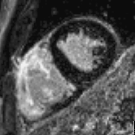
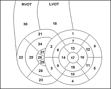
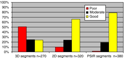
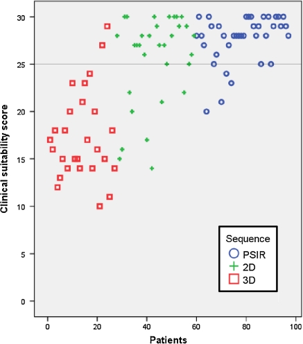
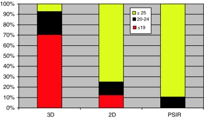
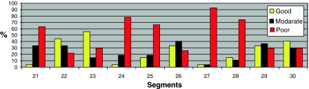
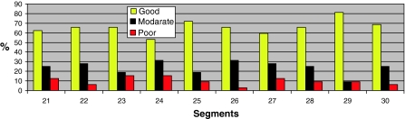
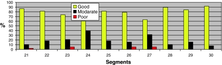
Histopathologic findings in arrhythmogenic right ventricular cardiomyopathy/dysplasia (ARVC/D) are replacement of the normal myocardium with fatty and fibrous elements with preferential involvement of the right ventricle. The right ventricular fibrosis can be visualised by post-gadolinium delayed enhancement inversion recovery imaging (DE imaging). We compared the image quality of three different gradient echo MRI sequences for short axis DE imaging of the right ventricle (RV). We retrospectively analysed MRI scans performed between February 2005 and December 2008 in 97 patients (mean age: 41.2 years, 67% men) suspected of ARVC/D. For DE imaging either a 2D Phase Sensitive (PSIR), a 2D (2D) or a 3D (3D) inversion recovery sequence was used in respectively 38, 32 and 27 MRI-examinations. The RV, divided in 10 segments, was assessed for image quality by two radiologists in random sequence. A consensus reading was performed if results differed between the two readings. Image quality was good in 24% of all segments in the 3D group, 66% in the 2D group and 79% in the PSIR group. Poor image quality was observed in 51% (3D), 10% (2D), and 2% (PSIR) of all segments. Exams were considered suitable for clinical use in 7% of exams in the 3D group, 75% of exams in the 2D group and 90% of exams of the PSIR group. Breathing-artifacts occurred in 22% (3D), 59% (2D) and 53% (PSIR). Motion-artifacts occurred in 56% (3D), 28% (2D) and 29% (PSIR). Post-gadolinium imaging using the PSIR sequence results in better and more consistent image quality of the RV compared to the 2D and 3D sequences.
Figures

References
-
- Basso C, Thiene G, Corrado D, et al. Arrhythmogenic right ventricular cardiomyopathy. Dysplasia, dystrophy, or myocarditis? Circulation. 1996;94(5):983–991. - PubMed
-
- Sen-Chowdhry S, Syrris P, Ward D, et al. Clinical and genetic characterization of families with arrhythmogenic right ventricular dysplasia/cardiomyopathy provides novel insights into patterns of disease expression. Circulation. 2007;115(13):1710–1720. doi: 10.1161/CIRCULATIONAHA.106.660241. - DOI - PubMed
-
- Maron BJ, Towbin JA, Thiene G, et al. Contemporary definitions and classification of the cardiomyopathies: an American Heart Association Scientific Statement from the Council on Clinical Cardiology, Heart Failure and Transplantation Committee; Quality of Care and Outcomes Research and Functional Genomics and Translational Biology Interdisciplinary Working Groups; and Council on Epidemiology and Prevention. Circulation. 2006;113(14):1807–1816. doi: 10.1161/CIRCULATIONAHA.106.174287. - DOI - PubMed
-
- Corrado D, Fontaine G, Marcus FI, McKenna WJ, et al. Arrhythmogenic right ventricular dysplasia/cardiomyopathy: need for an international registry. Study Group on Arrhythmogenic Right Ventricular Dysplasia/Cardiomyopathy of the Working Groups on Myocardial and Pericardial Disease and Arrhythmias of the European Society of Cardiology and of the Scientific Council on Cardiomyopathies of the World Heart Federation. Circulation. 2000;101(11):e101–e106. - PubMed
Publication types
MeSH terms
Substances
LinkOut - more resources
Full Text Sources
Other Literature Sources
Medical
