. 2011 Jul;49(7):599-618.
doi: 10.1002/dvg.20748. Epub 2011 Jun 21.

Isolation and live imaging of enteric progenitors based on Sox10-Histone2BVenus transgene expression

Affiliations

Isolation and live imaging of enteric progenitors based on Sox10-Histone2BVenus transgene expression

Jennifer C Corpening et al. Genesis. 2011 Jul.

Abstract

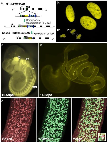
To facilitate dynamic imaging of neural crest (NC) lineages and discrimination of individual cells in the enteric nervous system (ENS) where close juxtaposition often complicates viewing, we generated a mouse BAC transgenic line that drives a Histone2BVenus (H2BVenus) reporter from Sox10 regulatory regions. This strategy does not alter the endogenous Sox10 locus and thus facilitates analysis of normal NC development. Our Sox10-H2BVenus BAC transgene exhibits temporal, spatial, and cell-type specific expression that reflects endogenous Sox10 patterns. Individual cells exhibiting nuclear-localized fluorescence of the H2BVenus reporter are readily visualized in both fixed and living tissue and are amenable to isolation by fluorescence activated cell sorting (FACS). FACS-isolated H2BVenus+ enteric NC-derived progenitors (ENPs) exhibit multipotency, readily form neurospheres, self-renew in vitro and express a variety of stem cell genes. Dynamic live imaging as H2BVenus+ ENPs migrate down the fetal gut reveals cell fragmentation suggesting that apoptosis occurs at a low frequency during normal development of the ENS. Confocal imaging both during population of the fetal intestine and in postnatal gut muscle strips revealed differential expression between individual cells consistent with down-regulation of the transgene as progression towards non-glial fates occurs. The expression of the Sox10-H2BVenus transgene in multiple regions of the peripheral nervous system will facilitate future studies of NC lineage segregation as this tool is expressed in early NC progenitors and maintained in enteric glia.


Figures

Figure 1
Figure 1. Sox10-H2BVenus BAC transgene construct and expression
a. Schematic diagram of Sox10-H2BVenus targeting vector and homologous recombination into the wild-type Sox10 BAC depicts Sox10 exons (black rectangles), initiator ATG (green), and Histone2BVenus (H2B) fusion reporter (grey/yellow). Sox10 homology arms (each approximately 500bp) are shown on either side of the Venus sequence. Excision of the TetR cassette after integration to derive the final Sox10-H2BVenus BAC transgene is shown. b. Expression of the H2BVenus reporter produces bright nuclear fluorescence in transfected enteric glial cells that is associated with mitotic chromosomes (insets b’ and b”) in a fraction of the transfected cells (400X). c. Sox10-H2BVenus transgene expression at 10.5dpc labels multiple neural crest derivatives (tg, trigeminal ganglia; ov, otic vesicle; v vagus; drg, dorsal root ganglion) in the PNS. d. Sox10-H2BVenus transgene in 14.5dpc fetal mouse gut labels enteric progenitors. e. Colocalization of Sox10 protein and the Sox10-H2BVenus transgene expression exhibits complete coincidence in fetal gut at 14.5dpc (400X). High magnification inset of a Sox10+/H2BVenus+ cell (white arrow) demonstrates the localization of the reporter protein with chromatin in mitotic spindles of dividing cells.
Figure 2
Figure 2. Appropriate temporal and spatial patterns of Sox10-H2BVenus transgene expression in migrating enteric NC-derived progenitors
a. Streams of H2BVenus+ cells are seen in whole-mount 9dpc Sox10-H2BVenus transgenic embryos imaged by confocal microscopy caudal to the otic vesicle in the glossopharyngeal (IX), vagal (X), and sympathetic chain pathways (sc) (200X). b. Subdissected 9dpc Sox10-H2BVenus transgenic gut reveals labeled ENP migrating into and down the developing foregut (200X). c. ENP progress into the midgut region of a whole-mount 10dpc gut (200X). Higher magnification inset shows heterogeneity of transgene expression at wavefront of migrating progenitors nearing the future cecal region (400X). Sacral NC labeled by Sox10-H2BVenus expression are indicated (arrow). d. Extent of H2BVenus+ ENP progress through the hindgut in a 12dpc sub-dissected transgenic embryo (200X). (ov, otic vesicle; ba1, brachial arch 1; ba2, brachial arch 2; ce, cecal region; hg, hindgut region; mg, midgut region)
Figure 3
Figure 3. Sox10-H2BVenus transgene expression is restricted to NC-derived enteric glia in the mature ENS
Gut muscle strips from adult Sox10-H2BVenus transgenic mice stained with antibodies to Sox10, S-100, PGP9.5 and Hu reveal expression of the transgene in mature enteric glia based on colocalization of the H2BVenus reporter with glial markers, Sox10 and S100. Sox10-H2BVenus expression is excluded from neurons labeled by PGP9.5 and Hu. All images 400X.
Figure 4
Figure 4. ENPs labeled by Sox10-H2BVenus expression exhibit stem cell characteristics
Sox10-H2BVenus+ cells from 14dpc fetal gut give rise to neurospheres in culture (a) and express the neural crest stem cell markers p75LNTR (b) and nestin (c) detected by Cy3 (red) IHC labeling (200X). Sox10-H2BVenus+ cells isolated from 14dpc gut grown in low density cultures give rise to multipotent colonies (d) that contain neurons (peripherin+), glia (GFAP+) and myofibroblasts (SMA+) labeled by triple immunofluorescence, 100X magnification. Detection of myofibroblasts with SMA-FITC resulted in co-visualization of SMA+ cells with H2BVenus+ nuclei in multipotent (SMA panel) colonies. RT-PCR detects expression (e) of stem cell genes (Abcg2, Bmi1, Dll1, Klf4, Lgr5, Msi1, Myc, Nes, Ngfr, Notch1, Pou5f1, Sox2), neural crest genes (FoxD3, Snai1, Snai2, Sox9, Sox10, Twist1), neuronal progenitor marker genes (Ascl1, Neurod1, Neurog1, Phox2b, Prph1, Uchl1), and peripheral glia markers (Erbb3, Fabp7, Gfap, Mpz, Nrg1, Nrtn, Olig2, S100b) in Sox10-H2BVenus+ enteric populations at discrete developmental stages. Housekeeping control genes (Actb, Ipo8, Ubc) are shown for comparison. Lanes include 100bp Molecular Weight Marker (M), no template control (H2O), total 14.5dpc fetal mouse RNA (Total E14), flow-sorted 14.5dpc gut H2BVenus+ ENPs (E14 Sox10+), cultured neurospheres from 14.5dpc H2BVenus+ gut (E14 NS), postnatal day 6 H2BVenus+ cells isolated from gut muscle strips (P6 GMS).
Figure 5
Figure 5. Sox10-H2BVenus transgene visualizes effects of Sox10Dom mutation on enteric development in situ
Enteric progenitors labeled by H2BVenus expression exhibit normal extent of migration in wild type 13.5dpc fetal guts in contrast to delayed migration in Sox10Dom/+ mutant littermates (a, 100X). High magnification confocal images (b, 400X) taken from wild type and Sox10Dom/+ mutant samples in gut regions labeled one to four in panel a at top to quantify proliferating progenitors. Proliferation of H2BVenus+ ENPs (white circles) is revealed by co-labeling with Sox10 antibody such that a red halo of Sox10 protein in the cytoplasm surrounds condensing H2BVenu+ mitotic chromosomes.
Figure 6
Figure 6. Proliferation of Sox10-H2BVenus+ ENPs observed by dynamic imaging of fetal gut
Nuclei of individual cells labeled by H2BVenus expression are apparent in selected frames of the hindgut from a 12.5dpc transgenic mouse. A single arrow highlights an ENP that undergoes cell division between 80 and 100 minutes of the recording with daughter cells indicated by two arrows. Other mitotic spindles are highlighted (asterisk). Scale bar, 20 microns. A complete frame series (Movie1) of this preparation is available at the Genesis website.
Figure 7
Figure 7. Elimination of Sox10-H2BVenus+ ENPs observed during dynamic imaging
Rapid fragmentation of cell nuclei that occur both at the wavefront (a) and behind the wavefront (b) are shown in selected frames of two different imaging sessions. Circles indicate the position of cell nuclei before (a, 100′; b, 280′), during, and after fragmentation. A complete frame series of the imaging session shown in panel b (Movie2) is available at the Genesis website. Nuclear fragmentation events were also identified in multiple independent fetal gut samples (d) that had been not illuminated prior to fixation and mounting as indicated by arrows. Scale bar panels a and b, 20 microns; scale bar panels shown in c, 10 microns.
Figure 8
Figure 8. Sox10-H2BVenus transgene labels NC-derived progenitors in multiple aspects of the PNS
Expression of the Sox10-H2BVenus transgene is apparent at 14.5dpc in other regions where NC contribute to developing structures including the cornea (a), follicles in the whisker pad (b), oligodendrocytes in the spine and glia of the dorsal root ganglia (c), nerve tracts in the forelimb (d) and innervation of the diaphragm (e).

References

    1. Abud HE, Young HM, Newgreen DF. Analysing tissue and gene function in intestinal organ culture. Methods Mol Biol. 2008;468:275–286. - PubMed
    1. Ahlstrom JD, Erickson CA. The neural crest epithelial-mesenchymal transition in 4D: a ‘tail’ of multiple non-obligatory cellular mechanisms. Development. 2009;136:1801–1812. - PMC - PubMed
    1. Andersen E, Asuri N, Clay M, Halloran M. Live imaging of cell motility and actin cytoskeleton of individual neurons and neural crest cells in zebrafish embryos. J Vis Exp. 2010 - PMC - PubMed
    1. Anderson RB, Stewart AL, Young HM. Phenotypes of neural-crest-derived cells in vagal and sacral pathways. Cell Tissue Res. 2006a;323:11–25. - PubMed
    1. Anderson RB, Turner KN, Nikonenko AG, Hemperly J, Schachner M, Young HM. The cell adhesion molecule l1 is required for chain migration of neural crest cells in the developing mouse gut. Gastroenterology. 2006b;130:1221–1232. - PubMed

Publication types

MeSH terms

LinkOut - more resources