

Glial vascular degeneration in CADASIL
- PMID: 21504125
- PMCID: PMC3019271
- DOI: 10.3233/jad-2010-100036
Glial vascular degeneration in CADASIL
Abstract
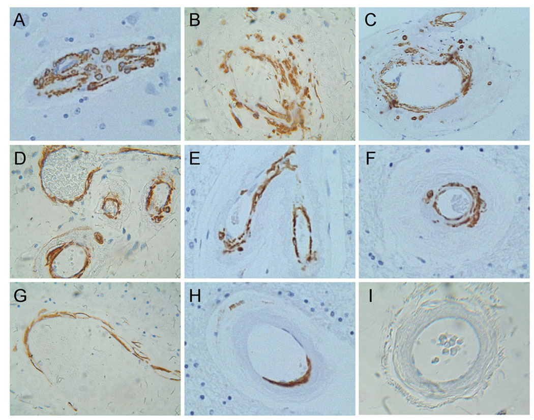
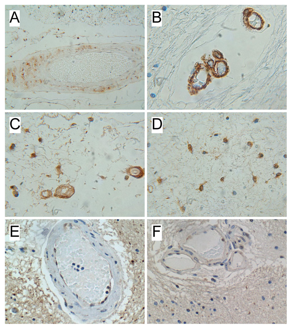
CADASIL is a genetic vascular dementia caused by mutations in the Notch 3 gene on Chromosome 19. However, little is known about the mechanisms of vascular degeneration. We characterized upstream components of Notch signaling pathways that may be disrupted in CADASIL, by measuring expression of insulin, IGF-1, and IGF-2 receptors, Notch 1, Notch 3, and aspartyl-(asparaginyl)-β-hydroxylase (AAH) in cortex and white matter from 3 CADASIL and 6 control brains. We assessed CADASIL-associated cell loss by measuring mRNA corresponding to neurons, oligodendroglia, and astrocytes, and indices of vascular degeneration by measuring smooth muscle actin (SMA) and endothelin-1 expression in isolated vessels. Immunohistochemical staining was used to assess SMA degeneration. Significant abnormalities, including reduced cerebral white matter mRNA levels of Notch 1, Notch 3, AAH, SMA, IGF receptors, myelin-associated glycoproteins, and glial fibrillary acidic protein, and reduced vascular expression of SMA, IGF receptors, Notch 1, and Notch 3 were detected in CADASIL-lesioned brains. In addition, we found CADASIL-associated reductions in SMA, and increases in ubiquitin immunoreactivity in the media of white matter and meningeal vessels. No abnormalities in gene expression or immunoreactivity were observed in CADASIL cerebral cortex. In conclusion, molecular abnormalities in CADASIL are largely restricted to white matter and white matter vessels, corresponding to the distribution of neuropathological lesions. These preliminary findings suggest that CADASIL is mediated by both glial and vascular degeneration with reduced expression of IGF receptors and AAH, which regulate Notch expression and function.
Figures
Similar articles
-
Is the increased expression of ubiquitin in CADASIL syndrome a manifestation of aberrant endocytosis in the vascular smooth muscle cells?J Clin Neurosci. 2008 May;15(5):535-40. doi: 10.1016/j.jocn.2007.06.022. Epub 2008 Mar 4. J Clin Neurosci. 2008. PMID: 18313300
-
Fibrosis and stenosis of the long penetrating cerebral arteries: the cause of the white matter pathology in cerebral autosomal dominant arteriopathy with subcortical infarcts and leukoencephalopathy.Brain Pathol. 2004 Oct;14(4):358-64. doi: 10.1111/j.1750-3639.2004.tb00078.x. Brain Pathol. 2004. PMID: 15605982 Free PMC article.
-
CADASIL: what component of the vessel wall is really a target for Notch 3 gene mutations?Neurol Res. 2004 Jul;26(5):558-62. doi: 10.1179/01610425016164. Neurol Res. 2004. PMID: 15265274 Review.
-
Severe white matter astrocytopathy in CADASIL.Brain Pathol. 2018 Nov;28(6):832-843. doi: 10.1111/bpa.12621. Brain Pathol. 2018. PMID: 29757481 Free PMC article.
-
[Cerebral autosomal dominant arteriopathy with subcortical infarcts and leukoencephalopathy (CADASIL)].Rinsho Byori. 2009 Mar;57(3):242-51. Rinsho Byori. 2009. PMID: 19363995 Review. Japanese.
Cited by
-
Mechanisms regulating cerebral hypoperfusion in cerebral autosomal dominant arteriopathy with subcortical infarcts and leukoencephalopathy.J Biomed Res. 2022 Aug 28;36(5):353-357. doi: 10.7555/JBR.36.20220208. J Biomed Res. 2022. PMID: 36165325 Free PMC article.
-
Recent Advances and Therapeutic Implications of 2-Oxoglutarate-Dependent Dioxygenases in Ischemic Stroke.Mol Neurobiol. 2024 Jul;61(7):3949-3975. doi: 10.1007/s12035-023-03790-1. Epub 2023 Dec 2. Mol Neurobiol. 2024. PMID: 38041714 Review.
-
Two novel mutations in NOTCH3 gene causes cerebral autosomal dominant arteriopathy with subcritical infarct and leucoencephalopathy in two Chinese families.Int J Clin Exp Pathol. 2015 Feb 1;8(2):1321-7. eCollection 2015. Int J Clin Exp Pathol. 2015. PMID: 25973016 Free PMC article.
-
Cell-to-cell communication and vascular dementia.Microcirculation. 2012 Jul;19(5):461-7. doi: 10.1111/j.1549-8719.2012.00175.x. Microcirculation. 2012. PMID: 22372561 Free PMC article. Review.
-
Dysfunction of Cerebrovascular Endothelial Cells: Prelude to Vascular Dementia.Front Aging Neurosci. 2018 Nov 16;10:376. doi: 10.3389/fnagi.2018.00376. eCollection 2018. Front Aging Neurosci. 2018. PMID: 30505270 Free PMC article. Review.
References
-
- Tournier-Lasserve E, Joutel A, Melki J, Weissenbach J, Lathrop GM, Chabriat H, Mas JL, Cabanis EA, Baudrimont M, Maciazek J, et al. Cerebral autosomal dominant arteriopathy with subcortical infarcts and leukoencephalopathy maps to chromosome 19q12. Nat Genet. 1993;3:256–259. - PubMed
-
- Joutel A, Corpechot C, Ducros A, Vahedi K, Chabriat H, Mouton P, Alamowitch S, Domenga V, Cecillion M, Marechal E, Maciazek J, Vayssiere C, Cruaud C, Cabanis EA, Ruchoux MM, Weissenbach J, Bach JF, Bousser MG, Tournier-Lasserve E. Notch3 mutations in CADASIL, a hereditary adult-onset condition causing stroke and dementia. Nature. 1996;383:707–710. - PubMed
-
- Chabriat H, Vahedi K, Iba-Zizen MT, Joutel A, Nibbio A, Nagy TG, Krebs MO, Julien J, Dubois B, Ducrocq X, et al. Clinical spectrum of CADASIL: a study of 7 families. Cerebral autosomal dominant arteriopathy with subcortical infarcts and leukoencephalopathy. Lancet. 1995;346:934–939. - PubMed
-
- Dichgans M, Mayer M, Uttner I, Bruning R, Muller-Hocker J, Rungger G, Ebke M, Klockgether T, Gasser T. The phenotypic spectrum of CADASIL: clinical findings in 102 cases. Ann Neurol. 1998;44:731–739. - PubMed
-
- Salloway S, Brennan-Krohn TSC, Mellion M, de la Monte S. CADASIL: A genetic model of arteriolar degeneration, white matter injury, and dementia in later life. In: Miller B, Boeve B, editors. Behavioral Neurology of Dementia. New York: Cambridge University; 2009. pp. 329–344.
Publication types
MeSH terms
Grants and funding
- RR15578/RR/NCRR NIH HHS/United States
- U01 AG032438/AG/NIA NIH HHS/United States
- R03 AG023916/AG/NIA NIH HHS/United States
- AA-12908/AA/NIAAA NIH HHS/United States
- R37 AA011431/AA/NIAAA NIH HHS/United States
- AA-11431/AA/NIAAA NIH HHS/United States
- P20 RR015578/RR/NCRR NIH HHS/United States
- R01 AA012908/AA/NIAAA NIH HHS/United States
- R56 AA011431/AA/NIAAA NIH HHS/United States
- AG023916/AG/NIA NIH HHS/United States
- R01 AA011431/AA/NIAAA NIH HHS/United States
- K24 AA016126/AA/NIAAA NIH HHS/United States
- K24-AA-16126/AA/NIAAA NIH HHS/United States
LinkOut - more resources
Full Text Sources
Miscellaneous
