Review
. 2011;17(13):1303-19.
doi: 10.2174/138161211795703735.

Drug discovery of antimicrobial photosensitizers using animal models

Affiliations
Review

Drug discovery of antimicrobial photosensitizers using animal models

Sulbha K Sharma et al. Curr Pharm Des. 2011.

Abstract

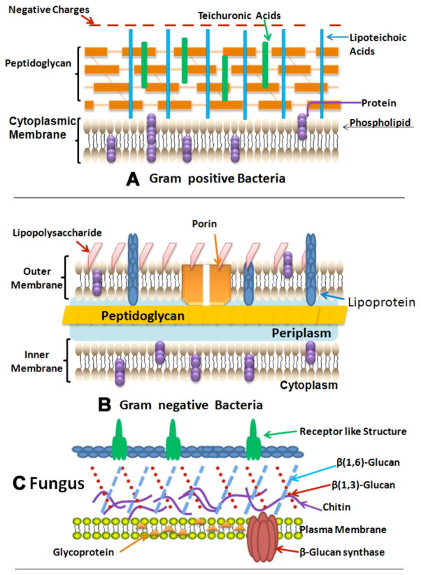
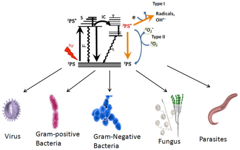
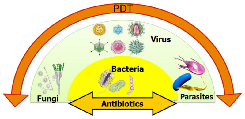
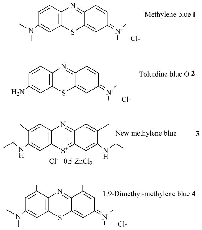
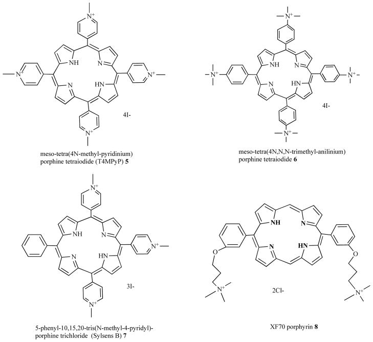
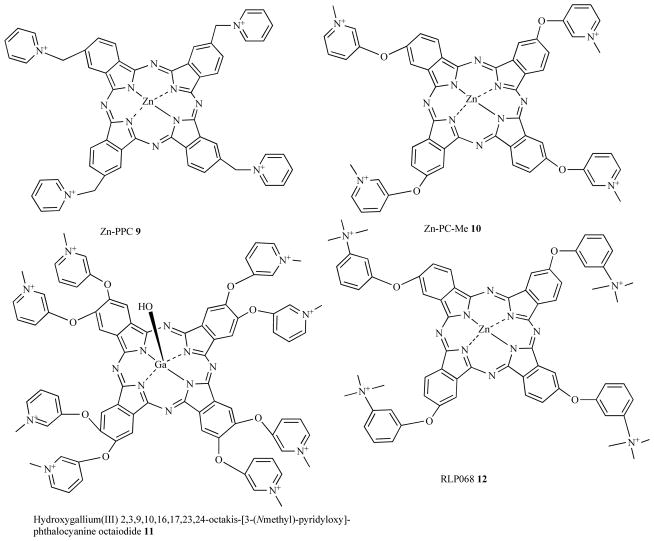
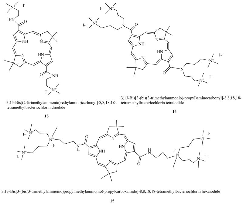
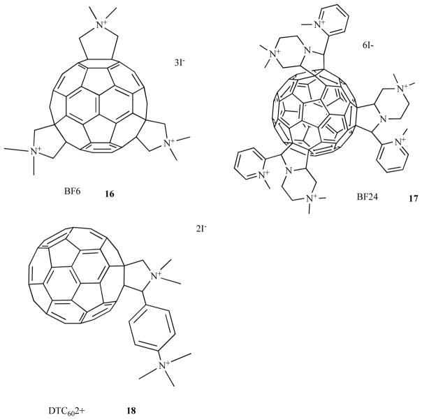
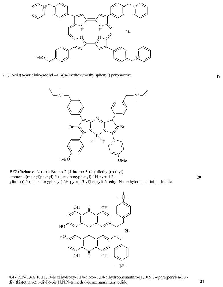
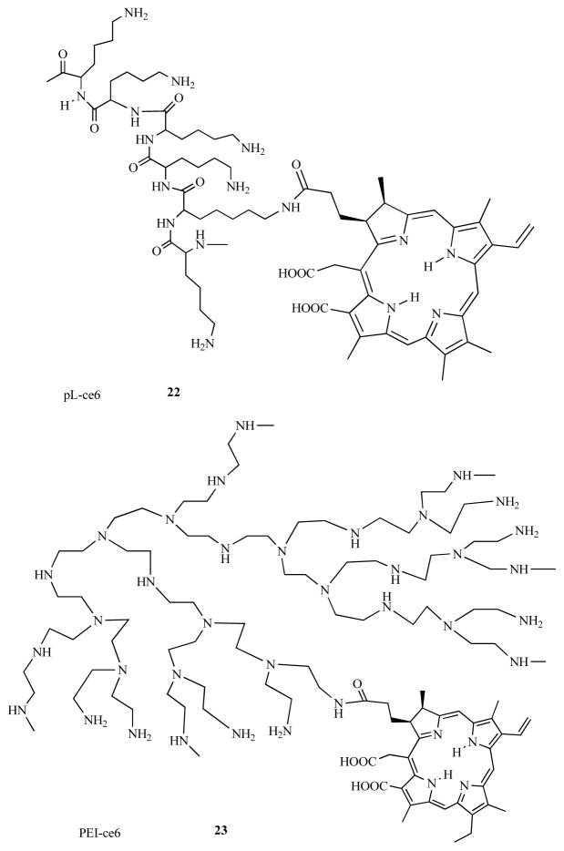
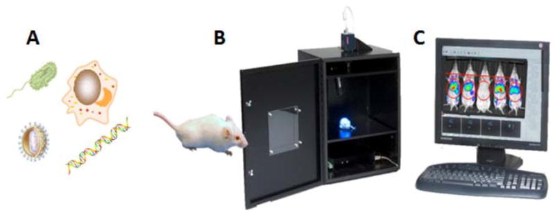
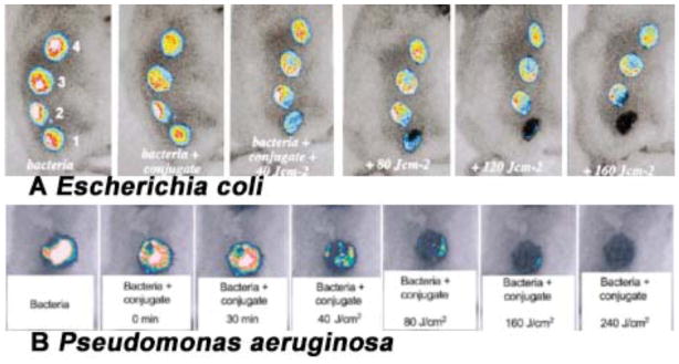
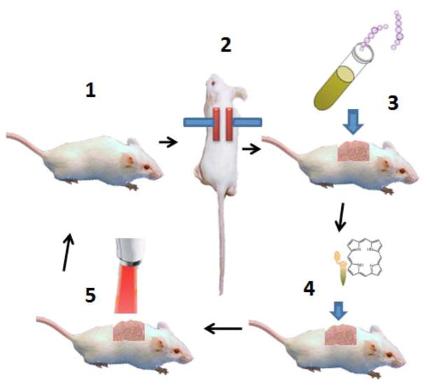
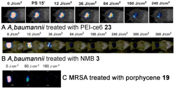
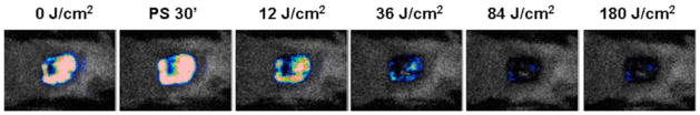
Antimicrobial photodynamic therapy (aPDT) is an emerging alternative to antibiotics motivated by growing problems with multi-drug resistant pathogens. aPDT uses non-toxic dyes or photosensitizers (PS) in combination with harmless visible of the correct wavelength to be absorbed by the PS. The excited state PS can form a long-lived triplet state that can interact with molecular oxygen to produce reactive oxygen species such as singlet oxygen and hydroxyl radical that kill the microbial cells. To obtain effective PS for treatment of infections it is necessary to use cationic PS with positive charges that are able to bind to and penetrate different classes of microbial cells. Other drug design criteria require PS with high absorption coefficients in the red/near infra-red regions of the spectrum where light penetration into tissue is maximum, high photostability to minimize photobleaching, and devising compounds that will selectively bind to microbial cells rather than host mammalian cells. Several molecular classes fulfill many of these requirements including phenothiazinium dyes, cationic tetrapyrroles such as porphyrins, phthalocyanines and bacteriochlorins, cationic fullerenes and cationic derivatives of other known PS. Larger structures such as conjugates between PS and cationic polymers, cationic nanoparticles and cationic liposomes that contain PS are also effective. In order to demonstrate in vivo efficacy it is necessary to use animal models of localized infections in which both PS and light can be effectively delivered into the infected area. This review will cover a range of mouse models we have developed using bioluminescent pathogens and a sensitive low light imaging system to non-invasively monitor the progress of the infection in real time. Effective aPDT has been demonstrated in acute lethal infections and chronic biofilm infections; in infections caused by Gram-positive, Gram-negative bacteria and fungi; in infections in wounds, third degree burns, skin abrasions and soft-tissue abscesses. This range of animal models also represents a powerful aid in antimicrobial drug discovery.


Figures

Fig. (1)
Fig. (1). Structures of the cell walls of three different classes of microbial pathogens
A) Gram-positive bacterium showing porous layer of peptidoglycan and single lipid bilayer. B) Gram-negative bacterium showing double lipid bilayer sandwiching peptidoglycan layer and an outer layer of lipopolysaccharide. C) Fungal cell with a less porous layer of beta-glucan and chitin surrounding a single lipid bilayer.
Fig. (2)
Fig. (2). Schematic mechanism of antimicrobial PDT
Type 1 and Type 2 photochemical mechanisms operate from photosensitizer triplet state producing ROS that are able to destroy all known microorganisms.
Fig. (3)
Fig. (3). Broad spectrum of effect of antimicrobial PDT
Many antibiotics, antibacterial and antifungal drugs have a relatively narrow spectrum of action, while antimicrobial PDT has an extremely broad spectrum of that takes effect rapidly.
Fig. (4)
Fig. (4). Structures of phenothiazinium dyes
Methylene blue, 1; Toluidine blue O, 2; New methylene blue, 3; Dimethyl-methylene blue, 4.
Fig. (5)
Fig. (5). Structures of cationic porphyrins
Tetra(4N-methyl-pyridyl) porphine tetraiodide (T4MPyP) 5, tetra(4N,N,N-trimethyl-anilinium) porphine tetraiodide (T4MAP) 6; 5-phenyl-10,15,20-tris(N-methyl-4-pyridyl)-porphine chloride (PTMPP or Sylsens B), 7; bis-cationic porphyrin, XF70, 8.
Fig. (6)
Fig. (6). Structures of cationic phthalocyanines
Tetrakis cationic zinc pyridinium phthalocyanine, Zn-PPC, 9; tetrakis cationic phthalocyanine 10; octakis-cationic Ga(III)-PC, 11; tetrakis cationic Zn-PC, RLP068, 12.
Fig. (7)
Fig. (7). Structures of cationic bacteriochlorins
Bis-cationic BC 13, Tetrakis-cationic BC 14, Hexakis-cationic BC 15.
Fig. (8)
Fig. (8). Structures of cationic fullerenes
Tris-methyl pyrrolidinium fullerene 16; hexakis-cationic fullerene, 17; bis-cationic fullerene, 18.
Fig. (9)
Fig. (9). Structures of miscellaneous cationic PS
Tris-cationic porphycene 19; bis-cationic brominated BF2 chelated tetraarylazadipyrromethene dye, 20; bis-cationic derivative of hypericin, 21.
Fig. (10)
Fig. (10). Structures of conjugates between PS and cationic polymers
Conjugate between poly-L-lysine and ce6, pL-ce6, 22; conjugate between polyethylenimine and ce6, PEI-ce6, 23.
Fig. (11)
Fig. (11). Schematic illustration of bioluminescence imaging to monitor PDT response in infection models
A) Different classes of pathogenic microorganisms have been rendered bioluminescent. B) Low light imaging camera consists of a light tight box with a sensitive CCD camera to collect emitted photons. C) Software allows the luminescence signal to be analyzed and the spread and intensity of the infection can be quantified over time.
Fig. (12)
Fig. (12). PDT for infected excisional wounds
(A) Successive overlaid luminescence images of a mouse with four excisional wounds infected with equal numbers of E. coli (5 ×10⁶). Wounds 1 (nearest tail) and 4 (nearest head) received topical application of pL-ce6 conjugate 22. Wounds 1 and 2 (two nearest tail) were then illuminated with successive fluences (40–160 J/cm2) of 665 nm light. (B) Successive overlaid luminescence false-color images of mice bearing excisional wounds infected with 5×10⁶ luminescent P. aeruginosa treated with pL-ce6 conjugate 22 and increasing doses of 660 nm light.
Fig. (13)
Fig. (13). Schematic illustration of procedures involved in carrying out PDT for burn infection
1. Shave and anesthetize a mouse. 2. Press two heated brass blocks against an elevated skin fold. 3. Add suspension of bioluminescent bacteria with a pipette tip. 4. Add photosensitizer with a pipette tip. 5. Deliver red light from a suitable light source.
Fig. (14)
Fig. (14)
A) PDT dose response of bacterial luminescence from a representative mouse burn infected with A. baumannii and treated with PDT using PEI-ce6 conjugate 23. B) PDT dose response of bacterial luminescence from a representative mouse burn infected with A. baumannii and treated with PDT using NMB 3. C) PDT dose response of bacterial luminescence from a representative mouse burn infected with MRSA and treated with PDT using PEI-ce6 conjugate 23.
Fig. (15)
Fig. (15). PDT for skin abrasion infected with MRSA
A) Successive bacterial luminescence images showing dose response with PDT using PEI-ce6 conjugate 23 of a representative mouse abrasion wound infected with luminescent MRSA. B) Successive bacterial luminescence images showing dark response with PEI-ce6 conjugate 23 of a representative mouse abrasion wound infected with luminescent MRSA. C). Time courses of bacterial luminescence of the infected abrasion wounds in the PDT treated mice (n=10) and non-treated mice (n=12). D) Kaplan-Meier wound healing curves of MRSA infected mouse abrasion wounds without treatment (neither PS nor light was applied) and treated with PDT, respectively.
Fig. (16)
Fig. (16). PDT of a Candida albicans infection
Dose response of fungal luminescence from a representative mouse skin abrasion wound infected with 106 CFU luminescent C. albicans and treated with new methylene blue 3 and 635-nm light at 24 hours after infection.
Fig. (17)
Fig. (17). Antimicrobial PDT is a rapidly growing field
The number of papers per year published in the general area of antimicrobial PDT obtained by a search of the MedLine database between 1954 and 2010.

Similar articles

Cited by

References

    1. Levy JG, Obochi M. New applications in photodynamic therapy. Introduction Photochem Photobiol. 1996;64:737–9. - PubMed
    1. Dougherty TJ, Gomer CJ, Henderson BW, Jori G, Kessel D, Korbelik M, Moan J, Peng Q. Photodynamic therapy. J Natl Cancer Inst. 1998;90:889–905. - PMC - PubMed
    1. Ochsner M. Photophysical and photobiological processes in the photodynamic therapy of tumours. J Photochem Photobiol B. 1997;39:1–18. - PubMed
    1. Raab O. Über die Wirkung fluoresceinder Stoffe and Infusorien. Zeit für Biol. 1900;39:524–546.
    1. Wainwright W. Photoantimicrobials - a PACT against resistance and infection. Drug Future. 2004;29:85–93.

Publication types